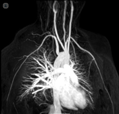
Magnetic resonance angiogram

Dr Paul Crowe - Interventional radiology
Created on: 12-29-2015
Updated on: 09-14-2023
Edited by: Aoife Maguire
What is magnetic resonance angiography?
Magnetic resonance angiography (MRA) is a technique which is used to evaluate and study the blood vessels and identify any abnormalities. A powerful magnetic field and radio frequency waves are used to produce detailed images of the arteries in the body on a computer screen. MRAs can examine blood vessels in the brain, neck, heart, chest, legs and feet, among other areas in the body.
What happens during a magnetic resonance angiography?
The patient lies on a table that slides into a large tunnel-shaped scanner. For this type of examination, a special dye is usually required as a contrast to visualise the areas with greater clarity. This contrast is almost always administered through a vein in the forearm or in the hand. During the test, the specialist radiologist observes the patient from another room for an hour or more.
Why is magnetic resonance angiography performed?
MRA is performed to examine the blood vessels of any part of the body (heart, head, abdomen, kidneys, lungs and legs) and thus detect diseases and conditions such as arterial aneurysm, aortic dissection, carotid artery disease, and stroke, among others.
Preparing for magnetic resonance angiography
Before the test, it is important to inform the specialist if you have any of the following:
- An artificial heart valve
- Clips for a cerebral aneurysm
- Implants in the inner ear (cochlear)
- A cardiac defibrillator or pacemaker
- An intrauterine device (IUD)
- A port of insulin or chemotherapy
- Neurostimulator
- Nephropathy or have received dialysis (possibly cannot receive contrast)
- A vascular stent
- Had artificial joints recently placed
It is also important to tell your doctor if you suffer from claustrophobia, as the test can take an hour or more. A medication to calm the anxiety may be administered in these cases.
You will be told not to eat or drink for 4-6 hours before the test. During the test, you will be given a hospital gown and asked to remove any clothing or accessories with metal clasps, since some types of metal can create blurry images. Therefore, carrying metal objects such as knives, glasses, credit cards, watches, jewelry, hearing aids, metal zippers, removable dental implants, among others, must be avoided.
What does the exam feel like?
The test does not present any kind of pain. The table can be cold and hard, but the patient can ask for a pillow. When the machine is turned on, it produces hums and thuds, so ear protectors may be provided. During the exam, you can talk to the person administering the test at any time thanks to an intercom in the room.
Meaning of abnormal MRA results
Abnormal MRA results can mean a problem in one or more blood vessels, among which are:
- Trauma
- Congenital disease
- Atherosclerosis
- Another vascular condition
Experts in Magnetic resonance angiogram
-

Dr Gavin Clague
RadiologyExpert in:
- MRI
- Musculoskeletal ultrasound
- Spine MRI
- Sports injuries
- Magnetic resonance angiogram
- CT scan (CAT)
- See all

Alliance Medical Cardiff
Alliance Medical Cardiff
G, Cardiff Gate Business Park Ltd, Copse Walk, Pontprennau CF23 8RB
No existe teléfono en el centro.
By using the telephone number provided by TOP DOCTORS, you automatically agree to let us use your phone number for statistical and commercial purposes. For further information, read our Privacy Policy
Top Doctors
-
Alliance Medical Cardiff
G, Cardiff Gate Business Park Ltd, Copse Walk, Pontprennau CF23 8RB, CardiffExpert in:
- Diagnostic Imaging
- Musculoskeletal imaging
- Magnetic resonance
- Spine MRI
- Rheumatology
- Most viewed diseases, medical tests, and treatments
- Cephalometric
- Medicolegal
- MRI
- OPG X-ray (Orthopantomography)
- CT scan (CAT)
- Sports injuries
- Proton beam therapy
- Port-a-cath insertion
- Nervous system malformations
- Brain MRI