Skin cancer detection: know your skin
Escrito por:Dr Hiba Injibar, a consultant dermatologist and founder of the Dermasurge clinic in London, has helped countless patients diagnose and manage skin cancer. As a specialist in skin cancer with over 20 years of experience, she shares with you her professional knowledge so that you know what to watch out for.
Understand your risk
Certain people are generally considered to have a higher risk of developing skin cancer. This includes people with pale skin, blue eyes, blonde hair and freckles. But remember, people of all skin and hair colours can develop skin cancer.
1. People with photosensitive skin
Another group who need to be extra vigilant are people on immunosuppressive medication – it makes their skin more photosensitive. Anyone with photosensitive skin can experience a reaction (e.g. a rash or solar urticaria ) when their skin is exposed to UV radiation from sunlight or tanning beds.
2. People with a family history of skin cancer
If you have a family history of skin cancer, you also need to be extra vigilant about your risk of the disease.
3. People often exposed to UV rays
If you work outside in the sun (therefore, exposing yourself to UV rays) for long periods, your risk of skin cancer is heightened. This is the same for people who use tanning beds. This heightened risk also extends to anyone who doesn’t use the appropriate SPF. If you know your skin will be exposed to UV rays, SPF is vitally important for protecting your skin.
Skin cancer detection: it’s not only about moles
Skin cancer can be very difficult to detect because cancerous skin doesn’t always lead to itchiness or pain. What’s more, it doesn’t only make itself known through moles. Watch out for slowly growing pimples or bumps that continue to grow. Any new skin lesion can signal the disease e.g. a growth, pimple or scaly patch.
We (dermatologists) highly recommend that you:
- get to know how your moles look (and regularly keep an eye on them).
- Check your skin for lesions (e.g. growths, pimples and scaly patches).
This includes checking the skin from difficult-to-see places e.g. your back and arms. To do this, we advise using a full-length mirror.
The ABCDE mnemonic
Many of you will have heard of the ABCDE mnemonic. We (and you at home) can use this mnemonic to check your risk.
Asymmetrical
Ask yourself if your mole is symmetrical. And, if it isn’t, ask yourself if the mole has always been asymmetrical.
Border
Is the border smooth around the edge or has it become irregular and jagged?
Colour
Consider if the colour of the mole has changed. Perhaps the mole overall is darker or pinkish, or maybe there are several new dark and/or pink patches within the mole.
Diameter
Is the mole growing larger in diameter? If the mole is growing, it needs checking. Particularly if the diameter is larger than 6 millimetres (about the size of the rubber at the end of a pencil).
Evolving
Taking all the above into consideration, is the mole evolving? Think about if the mole’s general appearance is different to how it was before.
Don’t hesitate to see a doctor
Make an appointment with your GP or a dermatologist right away if you detect a possible skin cancer symptom (like one of the criteria from the ABCDE mnemonic). If you visit your GP and they determine that there’s a cause for concern, they will very likely refer you to a dermatologist, such as myself, and then you will undergo an examination.
What to expect from your dermatological consultation
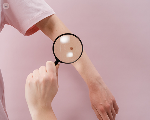
We use a special microscope (called a dermatoscope) to get an up-close and detailed look at your skin. It’s a handheld device that allows us to get a much more accurate view compared to what the human eye allows us to see.
With a dermatoscope, we can magnify the mole or skin lesion (remember, not only moles can suggest skin cancer) and we can evaluate the structure of the cells and how they are arranged. Once we get this up-close look at your skin, we’ll determine if we need to take a biopsy. Once the biopsy is analysed, we can determine the next step to take.
If skin cancer is not detected
If your results are negative for skin cancer, you may be asked to monitor your skin or you may be advised to have the mole/skin lesion removed as a precaution.
If you are diagnosed with skin cancer
After a skin cancer diagnosis, the mole is removed and you may be advised to undergo other cancer treatments. Your medical team will advise you on what your best options are.
Click here to learn how Dr Hiba Injibar can help you with the diagnosis and treatment of numerous skin conditions, including skin cancer.