Diagnosing prostate cancer: tests and surveillance
Autore:Prostate cancer is the most commonly diagnosed cancer in men - in fact it is thought most men will develop it if they live long enough. How can doctors diagnose prostate cancer? Expert urologist Mr Simon Bott is here with the answers.
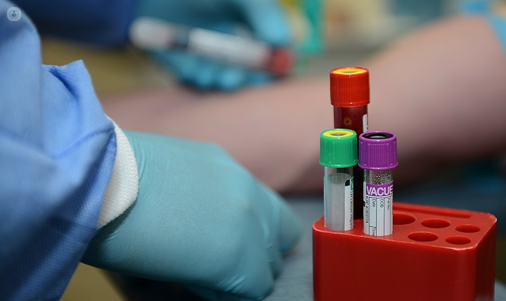
How is prostate cancer normally diagnosed?
Unfortunately, prostate cancer has to be quite advanced (in other words, it has to have grown quite a lot) in order for it to give you symptoms, and therefore we rely on a blood test called the PSA to detect that cancer before it's grown large and given rise to symptoms.
The PSA has some problems. Although it can diagnose prostate cancer early when the disease is very much curable, many men who don't have prostate cancer have a slightly high PSA. They would therefore be put through a number of investigations and it's reassuring for us to be able to tell them at the end that their results are negative, but the PSA may, in the meantime, have given them some anxiety.
The PSA is also very useful at monitoring treatment going forward – PSA is very reliable at telling us whether the cancer’s all gone or whether there are signs that it has come back.
If the PSA is high, we first check that you don't have any sign of infection, and if not, we then proceed to the next investigation, which would normally be an MRI scan. This looks inside the prostate to see if there's any abnormality, which could either be prostate cancer or something else that might be elevating the PSA.
What does being “under surveillance” mean?
We know that most men will develop prostate cancer if they live long enough and most men will never know about it in their lifetime. They will live a normal, full, healthy life, unaware they have prostate cancer.
Sometimes these prostate cancers are found either on a biopsy or an MRI scan and because we think they're never going to affect the patient during their normal lifetime, we recommend what's called active surveillance. This means we watch the patients very carefully with a combination of a blood test (the PSA), an MRI scan, and also sometimes biopsies to make sure the cancer isn't changing.
Sometimes, these cancers may show signs of change and then we'll then treat you with one of the normal therapies for prostate cancer, such as having your prostate removed, radiotherapy, brachytherapy or focal therapy.
The vast majority of people who start on active surveillance continue being monitored without the need for any form of treatment.
What are the challenges of making a diagnosis?
The challenges of making a diagnosis of prostate cancer include:
- The PSA – this doesn't always tell us definitively whether there's cancer there or not. The PSA can be raised by enlarged (but non-cancerous) prostates and urinary tract infections, so we look at these things to decide if there’s likely to be prostate cancer. If it is likely we do an MRI scan.
- Interpreting the MRI scan – while an MRI is much more accurate than a PSA test, or even a finger examination at the back passage, it doesn't always give us a black-and-white answer.
- Biopsy – if we think that the MRI shows us cancer we perform a biopsy. If the MRI is a bit equivocal (in other words it says there may or may not be cancer) we then make a decision based on the prostate size and the PSA as to whether or not a biopsy is necessary.
Often, the MRI is completely normal and therefore you wouldn't need to have any biopsy at all but maybe carry on watching your blood tests going forward just to check.
How do fusion-guided biopsies improve the process?
MRI is fantastic at looking inside the prostate to see if there is any prostate cancer there. When we do a biopsy, we unfortunately can't do this using an MRI scanner to identify exactly where the tumour is. An MRI scanner is a noisy tunnel and it would be difficult for the urologist to get in to access the prostate.
To solve this problem, we do an ultrasound scan by placing a small probe up the backside. Ultrasound is very good at showing where the prostate is and the size and shape of the prostate, but it doesn't give detail about what's going on right inside the prostate.
Fusion biopsies are a recent development which outlines the prostate and the abnormal area within it on the MRI scan and then fuses those outlines with the ultrasound image. This enables the urologist to see on the ultrasound image the abnormalities that we've detected on the MRI scan. What I can then do with a fusion biopsy is put a needle directly into what is seen on the MRI scan using the ultrasound scan, so I know I've hit the right target.
The fusion biopsy then also records exactly where I've taken the biopsy, so we have a record going forward of exactly where that abnormality is and where the biopsy came back positive or negative for prostate cancer as this can impact treatment going forward.