Phacoemulsification cataract surgery: what you need to know
Written by:There are different methods ophthalmologists use to remove cataracts during surgery. One method is phacoemulsification, an ultrasound technique performed through a micro incision. Mr Sivanandy Nagendran, an ophthalmologist and cataract specialist, exlplains this surgery and what to expect before, during, and after.

What is phacoemulsification cataract surgery?
Phacoemulsification surgery is the modern surgical technique using ultrasound to remove the cloudy part of the lens through a micro incision. After removal of the cataract, a clear lens is inserted in to your eye. Practically all cataracts are removed using this method and each procedures takes 10 to 15 minutes to complete. Over 95 percentage of phacoemulsification surgery in the UK is done under local anaesthesia as a day case procedure.
What happens before the cataract surgery?
An optician often diagnoses a cataract during one of your regular eye tests. If they think that your sight cannot be corrected with new spectacles or if your symptoms affect your quality of life, they will refer you to see an ophthalmologist.
Your ophthalmologist will examine your eyes to assess the severity of the cataract and to make sure the rest of your eye is healthy. This may include dilating your pupils with drops. They also take certain measurements of your eyes to calculate the strength of the implant needed.
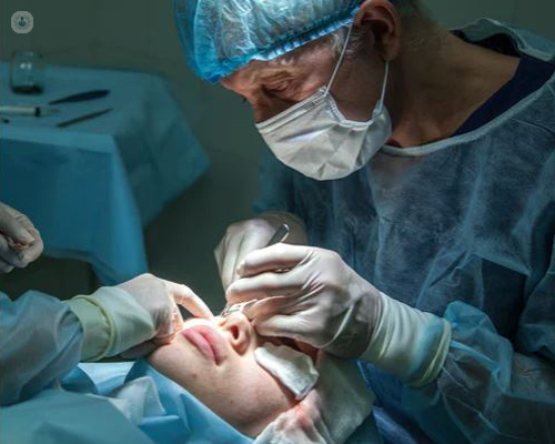
What happens during surgery?
Cataract surgery is carried out in a sterile operating theatre. Before the surgery, you will be given drops to dilate the pupils to get a clear view of the cataract. Your eye is then anaesthetised using eye drops and sometimes with additional local anaesthetic injection around the eye. A very small group of patients need general anaesthetic.
Your eye and the surrounding area will then be cleaned with antiseptic fluid. In order to protect from infection, your face will then be covered with a sterile sheet so that only the eye being operated on is exposed. Once your eye is numb your ophthalmologist will start the procedure. Throughout the procedure someone will be holding your hand to keep you comfortable.
Your ophthalmologist will make some very small cuts through the cornea, which allows them to introduce instruments through your dilated pupil to reach your lens.
During the surgery you may be able to see movement and a change in lights or shadows, but it’s unlikely that you will be able to see any detail of what’s happening. You also will hear noise made by the ultrasound machine.
The lens in your eye is made up of different layers, and the outside layer is called the lens capsule. During the operation, the ophthalmologist makes a circular opening through the front of the lens capsule so that they can reach the lens inside. Using the same instrument, the ophthalmologist can break up your cloudy lens and remove it using suction. Your lens capsule is kept in place so that the artificial lens implant can be placed inside it. The tiny implant is folded so that it can be put into your eye through the same tiny incision that has been used to remove your cataract. The lens unfolds within the eye and is held in place by the lens capsule. These small incisions are self-sealing and usually do not need suturing.
At the end of the operation your eye may be covered with a shield to keep it clean and for protection. Then, once your ophthalmology team is happy with your eye, you’ll be able to go home. You will be prescribed with anti-inflammatory and antibiotic eye drops.
What are the results like?
Cataract surgery offers good long-term results, and more than 97 per cent of all cases done by an experienced ophthalmologist are successful and free of complications. Therefore, the risks of getting serious complications are very rare. The most common complications can be treated with eye drops or further surgery. There is a very small risk—around 1 in 1000—of permanent sight loss in a treated eye as a direct result of the procedure.