What does a pathologist examine?
Written by:A pathologist examines cells, tissues and body fluid with the use of a microscope or with other diagnostic tools. Have you been referred to a pathologist and wondered what they will analyse, how they do it and why? Dr Ashish Chandra explains exactly what he does as a pathologist, who specialises in histopathology, cytology and fine needle aspiration.
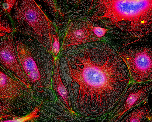
Pathologies: what's the difference between histopathology and cytopathology?
My job involves making a diagnosis and checking for infection, disease, cancer or precancerous conditions, by looking down the microscope at tissues or cells. When I make a diagnosis on tissues, this is called histopathology and in particular, I have considerable experience in urological histopathology which involves diseases of the kidney, the bladder, the prostate or the testes.
Cytopathology is the study of cells and making a diagnosis on the basis of the cellular appearances of an organ or a tissue under the microscope. This can be done in one of many ways. I could be sent some slides to review either histopathology or cytopathology, which may have been taken at another pathology lab at another hospital. These could simply be sent across to me and I would provide a second opinion for your physician and be available to discuss the results with them if they wish to do so.
When would I need fine needle aspiration (FNA)?
I also perform fine needle aspiration, which is a diagnostic procedure where I would examine and look at where small lumps were felt in the neck or any other parts of the body. A physician may have referred you to me if they found lumps whilst examining you and would like a needle test to be performed to ascertain the nature of that lump.
This procedure involves the use of a very small and very fine needle, for which no local anaesthetic is necessary although a cream or an ointment is available to apply before the procedure so that the skin is numb. The needle pinprick is not felt quite as much.
Alternatively, I may call upon the assistance of my radiologist colleagues if the lump is very small or difficult to feel. This may involve the use of actually injecting a local anaesthetic into the skin provided you’re not allergic to it and have not had any reactions in the past.
This allows us to take multiple samples, very small samples, using either a fine needle or a core biopsy needle. This would be examined either on the spot or taken back to the laboratory for additional tests to be performed. I would provide you with an assessment of the adequacy of the sample on the spot and let you know whether enough cells have been collected for an adequate diagnosis and for the tests to be performed.
What other methods does a pathologist use?
The additional tests that sometimes need to be performed are flow cytometry, which is a laser-based technology used if a lymphoma is suspected, or immunochemistry to ascertain the origin of a tumour or genetic and molecular tests on a range of tumours.
If you think you need a consultation with a pathologist for either cytology, fine needle aspiration cytology or histological pathology involving the kidney, bladder, prostate or testes, then please book an appointment to see me.