Electromyography

Dr Taimour Alam - Neurophysiology
Created on: 07-12-2013
Updated on: 06-01-2023
Edited by: Conor Lynch
What is an electromyography?
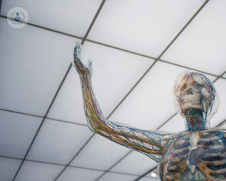
An electromyography (EMG) is a diagnostic test to analyse the health of the muscles and the nerve cells (neurons) that control them by analysing electrical activity in the muscles.

What does a electromyography involve?
Electromyography consists of two parts: a nerve conduction study and a needle EMG. The nerve conduction study involves putting electrodes on the skin in the area that has been experiencing symptoms. These measure the speed and strength of electrical signals between different points to evaluate how well the motor neurons communicate with the muscles.
In the needle EMG, electrodes are inserted into the muscle via a needle. These evaluate electrical activity within the muscle tissue both when the muscle is contracted and when it is at rest. After the test is complete, the electrodes are removed.
The readings from the electrodes are sent to a computer, which displays them as graphs and/or numerical values, which your doctor can interpret for you.
Why is electromyography done?
Electromyography is used to diagnose or rule out a number of conditions that affect the nerves and/or muscles. A doctor may order an EMG if the patient presents with the following symptoms:
- Muscle weakness
- Tingling
- Numbness
- Cramps
- Paralysis
- Limb pain
- Involuntary twitching (tics)
What does the test feel like?
You may be asked to lie or sit down during an electromyography. Before the needle EMG, the doctor will clean the area where the needle is to be inserted with antiseptic. You may feel some pain or discomfort when the needle is inserted.
What do abnormal results mean?
If electromyography yields abnormal results, it could indicate:
- Muscle disorders, e.g. muscular dystrophy, polymyositis
- Disorders that affect the connection between the motor neuron and the muscle, e.g. myasthenia gravis
- Peripheral nerve (nerves outside the spinal cord) disorders, e.g. carpal tunnel syndrome, peripheral neuropathy
- Conditions affecting the motor neurons in the spinal cord or brain, e.g. polio, amyotrophic lateral sclerosis (ALS)
- Conditions that affect the nerve root, e.g. herniated spinal disc

An expert guide: Nerve conduction studies and EMG
By Dr Taimour Alam
2024-04-25
In this informative guide for patients, revered consultant neurophysiologist Dr Taimour Alam shares his expert insight on nerve conduction and EMG studies. See more
Experts in Electromyography
-
Dr Taimour Alam
NeurophysiologyExpert in:
- Carpal tunnel syndrome
- Electromyography
- Peripheral neuropathy
- Neuromuscular disease
- Diabetic neuropathy (diabetes pain)
- Medicolegal
- See all
- Most viewed diseases, medical tests, and treatments
- Medicolegal
- Sleep disorders
- Carpal tunnel syndrome
- Doppler Ultrasound
- Transcranial magnetic stimulation (TMS)
- Restless legs syndrome
- Epilepsy
- Pelvic pain
- Insomnia
- Adult ADHD