Magnetoencephalography

What is magnetoencephalography?

Magnetoencephalography (MEG) is a medical test used to analyse brain activity by measuring the magnetic fields produced by electrical currents in the brain. It is arguably the most advanced method of recording and analysing a functioning brain, holding advantages over similar complementary techniques.

Magnetoencephalography testing can measure brain activity by the millisecond, giving it an advantage over MRI scans, and can determine where in the brain activity is occurring with greater accuracy than an EEG (electroencephalogram).
What does magnetoencephalography consist of?

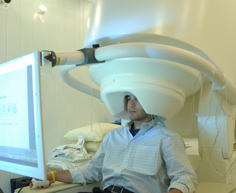
Magnetoencephalography involves using special equipment to measure the small magnetic fields generated by neurons firing in the brain. The scan utilises a superconducting quantum interference device (SQUID) and a computer to measure this neuromagnetic activity and superimposes the results over an anatomical picture of the brain.

The SQUID is highly sensitive and is housed in a magnetically shielded room to negate interference from the Earth’s magnetic field, which is a billion times stronger than those generated by the brain.
 

Why is magnetoencephalography done?

Magnetoencephalography is done to map brain function for diagnostic reasons. One particular use is to identify the source of epileptic seizures within the brain.

It is also used in preoperative and pre-treatment planning for individuals with epilepsy, brain tumours, or other lesions. MEGs are also useful in research, helping scientists to understand the brain.
 

Preparation for magnetoencephalography

Jewellery and accessories are not allowed into the room, as they can interfere with the test, so it may be best to leave such items at home. Patients with implants (particularly metal ones) may not be able to have the scan for the same reason.

As with all medical tests and procedures, follow your doctor’s instructions both before and during the test.
 

What to expect during the test

Magnetoencephalography is non-invasive and usually performed as a day case. The patient sits in a magnetically-shielded room, and a large helmet full of magnetic sensors is placed on the patient’s head. It fits loosely, so claustrophobia is rarely an issue.

An EEG may be performed at the same time, which will involve sticking electrodes to the patient’s scalp.

You may have to perform certain actions, answer questions, look at images, listen to sounds, or read to assess how you respond to various stimuli and identify which parts of the brain are responsible for different things. You may also have to sleep during the test.

08-10-2023
Top Doctors

Magnetoencephalography

Dr Taimour Alam - Neurophysiology

Created on: 07-12-2013

Updated on: 08-10-2023

Edited by: Sophie Kennedy

What is magnetoencephalography?

Magnetoencephalography (MEG) is a medical test used to analyse brain activity by measuring the magnetic fields produced by electrical currents in the brain. It is arguably the most advanced method of recording and analysing a functioning brain, holding advantages over similar complementary techniques.

Magnetoencephalography testing can measure brain activity by the millisecond, giving it an advantage over MRI scans, and can determine where in the brain activity is occurring with greater accuracy than an EEG (electroencephalogram).

What does magnetoencephalography consist of?

Magnetoencephalography involves using special equipment to measure the small magnetic fields generated by neurons firing in the brain. The scan utilises a superconducting quantum interference device (SQUID) and a computer to measure this neuromagnetic activity and superimposes the results over an anatomical picture of the brain.

The SQUID is highly sensitive and is housed in a magnetically shielded room to negate interference from the Earth’s magnetic field, which is a billion times stronger than those generated by the brain.
 

Why is magnetoencephalography done?

Magnetoencephalography is done to map brain function for diagnostic reasons. One particular use is to identify the source of epileptic seizures within the brain.

It is also used in preoperative and pre-treatment planning for individuals with epilepsy, brain tumours, or other lesions. MEGs are also useful in research, helping scientists to understand the brain.
 

Preparation for magnetoencephalography

Jewellery and accessories are not allowed into the room, as they can interfere with the test, so it may be best to leave such items at home. Patients with implants (particularly metal ones) may not be able to have the scan for the same reason.

As with all medical tests and procedures, follow your doctor’s instructions both before and during the test.
 

What to expect during the test

Magnetoencephalography is non-invasive and usually performed as a day case. The patient sits in a magnetically-shielded room, and a large helmet full of magnetic sensors is placed on the patient’s head. It fits loosely, so claustrophobia is rarely an issue.

An EEG may be performed at the same time, which will involve sticking electrodes to the patient’s scalp.

You may have to perform certain actions, answer questions, look at images, listen to sounds, or read to assess how you respond to various stimuli and identify which parts of the brain are responsible for different things. You may also have to sleep during the test.

This website uses our own and third-party Cookies to compile information with the aim of improving our services, to show you advertising related to your preferences as well analysing your browsing habits. You can change your settings HERE.