Ultrasound

Dr Marina Fernando - Obstetrics & gynaecology
Created on: 11-13-2012
Updated on: 11-07-2023
Edited by: Sophie Kennedy
What is an ultrasound?
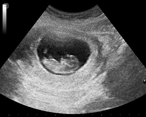
Ultrasound is a diagnostic imaging procedure that allows you to see organs and soft structures inside the body. At present, they can be 2D, 3D or four-dimensional images. Ultrasound is a non-invasive technique, in which radiation is not used. Thanks to this, it is used to visualise developing foetuses in pregnancy.
What does an ultrasound scan consist of?
It is carried out using high frequency sound waves emitted through a transducer that captures the echo of different amplitudes which occurs when they bounce off the organs. These signals, once processed by a computer, give a series of images.
Why are ultrasound scans carried out?
With this procedure you can clearly differentiate the shape and size of each structure inside the body. In medicine it is used to see the heart, kidneys, liver and blood vessels, among other organs. In addition, ultrasounds are also carried out on patients who are pregnant.
Another function of this ultrasound procedure is the study of different tissues of the body such as the blood flow of arteries and veins to detect arteriosclerosis and blood clots. Ultrasounds also allow the analysis of the thyroid gland and other soft structures of the neck as well as tendons, ligaments, muscles and joint structures.
How do you prepare for an ultrasound scan?
Usually, you do not need any type of preparation for having an ultrasound. In cases where the area to be examined is the abdomen, the specialist may tell you to drink three glasses of water approximately one hour before the test.
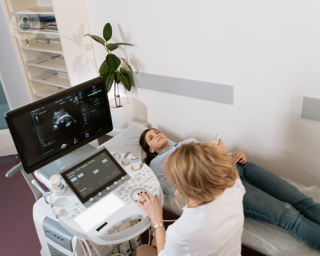
What happens during an ultrasound scan?
During the ultrasound, the patient will lie on a table or stretcher with the area to be analysed exposed. The specialist will apply a transparent gel that will allow the movement of the roller built into the transducer. During the scan, the expert will interpret the images and determine which shots are correct according to each case.
The only slight discomfort that can be felt is the gentle pressure that the specialist applies when passing the transducer over the body area. The gel applied can also feel quite cold when applied.
Abnormal ultrasound results
The types of images that are considered abnormal are:
- congenital anomalies
- an alteration to tissue due to trauma or injury
- cysts


All about the percutaneous nephrolithotomy procedure
By Mr Ivo Dukic
2024-04-17
For individuals diagnosed with large or complex kidney stones, navigating treatment options can be overwhelming. Percutaneous nephrolithotomy (PCNL) is a minimally invasive approach offering greater stone clearance compared to alternative methods. Mr Ivo Dukic, leading consultant urological surgeon, explains the PCNL procedure including exciting advancements in miniaturisation of this key hole procedure in this in-depth article. See more

What are the different types of ultrasound scans in pregnancy?
By Dr Srividhya Sankaran
2024-04-17
Different types of ultrasound are undertaken depending on the stage of the pregnancy, and also for various reasons. These can include calculating the chances of the pregnancy being affected with Down’s syndrome and risk of going into preterm labour, alongside many others. Highly-experienced consultant in maternal-foetal medicine (MFM) and obstetrics Dr Srividhya Sankaran, is here to explain these different ultrasound scans. See more


An expert's guide to third trimester ultrasounds
By Dr Spyros Bakalis
2024-04-16
Third trimester ultrasounds are an important step in pregnancy, reassuring the mother that her baby is growing healthily. In his latest online article, highly-experienced consultant in obstetrics and maternal and foetal medicine Dr Spyros Bakalis explains this scan in detail. See more


Understanding the difference between a miscarriage and an ectopic pregnancy, and where to get treatment
By Dr Shahla Ahmed
2024-04-16
Ectopic pregnancies and miscarriages have similar symptoms, making it difficult to differentiate between them without seeing a specialist. Dr Shahla Ahmed, a leading consultant gynaecologist in London, explains what these conditions are, how you can tell them apart and where to get treated. See more
Experts in Ultrasound
-

Mr Keith Duncan
Obstetrics & gynaecologyExpert in:
- Breakthrough bleeding
- Caesarean
- Childbirth
- Multiple pregnancy
- Ultrasound
- High-risk pregnancy
-

Dr Ralph Rogers
Sports medicineExpert in:
- Sports injuries
- Platelet-rich plasma
- Back pain
- Lipogems
- Knee osteoarthritis
- Ultrasound
-

Dr Paul Crowe
Interventional radiologyExpert in:
- Fibroids
- Pelvic congestion syndrome
- Varicocele
- Ultrasound
- Prostate artery embolisation
- CT scan (CAT)
-

Dr Sharmistha Guha
Obstetrics & gynaecologyExpert in:
- Pregnancy counselling
- Ultrasound
- Minimal access surgery (keyhole surgery)
- Colposcopy
- Endometriosis
- Menstrual disorders
-
Mr Ahmad Sayasneh
Obstetrics & gynaecologyExpert in:
- Endometriosis
- Heavy periods
- Pelvic pain
- Gynaecological cancer
- Colposcopy
- Ultrasound
- See all

The Princess Grace Hospital - part of HCA Healthcare
The Princess Grace Hospital - part of HCA Healthcare
The Princess Grace Hospital, 42-52 Nottingham Pl
No existe teléfono en el centro.
By using the telephone number provided by TOP DOCTORS, you automatically agree to let us use your phone number for statistical and commercial purposes. For further information, read our Privacy Policy
Top Doctors
Spire Little Aston Hospital
Spire Little Aston Hospital
Little Aston Hall Drive, Sutton Coldfield, B74 3UP
No existe teléfono en el centro.
By using the telephone number provided by TOP DOCTORS, you automatically agree to let us use your phone number for statistical and commercial purposes. For further information, read our Privacy Policy
Top Doctors
Guy’s and St Thomas’ Private Healthcare
Guy’s and St Thomas’ Private Healthcare
Guy’s Hospital, Great Maze Pond
No existe teléfono en el centro.
By using the telephone number provided by TOP DOCTORS, you automatically agree to let us use your phone number for statistical and commercial purposes. For further information, read our Privacy Policy
Top Doctors
-
The Princess Grace Hospital - part of HCA Healthcare
The Princess Grace Hospital, 42-52 Nottingham Pl, Central LondonExpert in:
- Cancer
- General Surgery
- Orthopaedic surgery
- Robotic Surgery
- Intensive care
- Sports Medicine
-

Spire Little Aston Hospital
Little Aston Hall Drive, Sutton Coldfield, B74 3UP, Sutton ColdfieldExpert in:
- Bariatric Surgery
- General Surgery
- Orthopaedic surgery
- Robotic Surgery
- Diagnostic Imaging
- Ophthalmology
-

Guy’s and St Thomas’ Private Healthcare
Guy’s Hospital, Great Maze Pond, SE1 South Bank LondonExpert in:
- Allergy
- Cardiology
- General Surgery
- Maxillofacial Surgery
- Thoracic Surgery
- Maternity care
- See all
- Most viewed diseases, medical tests, and treatments
- Anxiety
- Family history of breast cancer
- Digestive diseases
- Stress
- Women's health
- Cephalometric
- Hypertension (high blood pressure)
- Medicolegal
- Endometriosis
- MRI