Advantages of an endoscopic ultrasound
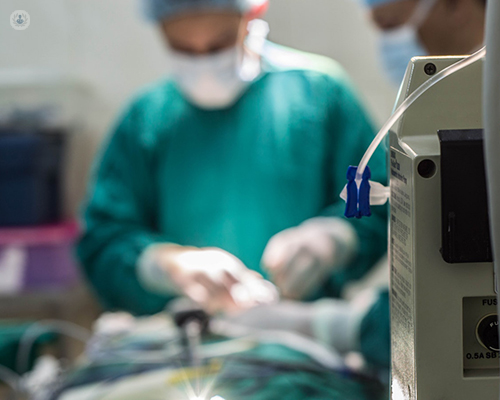
Escrito por:Endoscopy is a department where specialist equipment or cameras (endoscopes) are passed directly inside the human body. These cameras can be passed into the throat, lungs, stomach, small bowel, bile ducts, large bowel and bladder by specially trained medical professionals or endoscopists.
An endoscopic ultrasound (EUS) is a procedure where a specially adapted ultrasound probe is placed on the end of an endoscope to obtain high-quality images of the organs inside the body. Expert gastroenterologist Mr Mark Austin explains when a EUS is necessary and the advantages of the procedure.
How does endoscopic ultrasound work?
The endoscope is passed through the mouth and gullet, into the stomach and small bowel, as in a normal gastroscope examination. As the endoscope is slightly larger than a standard scope and the fact that the procedure takes slightly longer, sedation is always used.
What is the purpose of an endoscopic ultrasound?
When the ultrasound probe is placed within the body, it allows very detailed pictures of specific structures or organs deep inside. The area in question can then be seen and assessed with special software in the ultrasound processing unit or scanner. This software allows for the determination of the position of local blood vessels, whether the lesion is solid or liquid or whether there are characteristics seen with tumours or cancers. Importantly, the lesion or area can then be sampled with a needle passed through the scope. This piece of tissue is sent to the laboratory allowing for analysis and to aid diagnosis.
What are the risks of this procedure?
This endoscopic procedure carries very little risk to patients, without any exposure to X-rays or radiation. The risks can be very simple from a sore throat or bloating to more serious complications, which obviously depend on what intervention is performed.
What are the advantages of an endoscopic ultrasound?
The benefits of this procedure over standard abdominal ultrasound scan, MRI or CT scan are well recognised and it is used preferentially to gain tissue and accurately understand the particular stage of cancer. The organs that can be seen and easily sampled include the oesophagus or gullet, lymph nodes, the stomach, liver, adrenals and pancreas gland. It is very useful for confirming gallstone disease, underlying liver disorders and pancreatic abnormalities including cancer. The procedure lasts about 30 minutes and you will receive a report at the end informing you of all the results.
Do not hesitate to book an appointment with a specialist.