Under the microscope: the world of histopathology
Escrito por:Diagnosing an illness is not always a simple task. It can take a team of specialists working together to find the answer and offer up a solution. Sometimes, samples of the patient’s tissue may be taken to be analysed in a laboratory, and this is where the fascinating field of histopathology comes into play. We asked Dr Morgan Moorghen about the life and work of a histopathologist.
What is histopathology?
Histopathology is the study of diseases based on the examination of tissues under a microscope. Histopathologists are therefore doctors who make diagnoses based on the examination of small biopsies and also larger specimens retrieved at an operation performed by a surgeon. Many histopathologists specialise in diseases of specific organs such as the bowel, the lungs etc. Practically all cancers are diagnosed by histopathologists and the information provided in the histopathology report is crucial in formulating the correct treatment plan for the patient.
The histopathologist within a multidisciplinary team
The management of patients with complex diseases such as cancer requires the input of a range of professionals, including specialist nurses, surgeons, oncologists, radiologists, physicians and histopathologists who constitute the multidisciplinary team (MDT). The multidisciplinary team meeting (MDTM) is the forum which allows for the diagnosis and management of each patient to be discussed in order to optimise clinical outcome. At the MDTM, the histopathologist has an important role in providing the diagnosis and discussing aspects of treatment prognosis based on microscopic features elucidated in the tissue.
The regular working day of the histopathologist
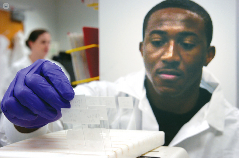
The main task of the histopathologist is to examine tissue slides under a microscope. It is estimated that in the UK some 20 million slides are examined annually. For each biopsy or other specimen received in a histopathology laboratory, the number of slides which are generated for examination by the histopathologist varies between 1 and in excess of 100 for the more complex cases.
Having examined the slides, the histopathologist would normally dictate a report, which is then transcribed and uploaded on the computer system and made available to the members of the team who are looking after the patient.
The other important task for a histopathologist relates to the examination and dissection of the larger specimens (e.g. a bowel specimen which contains cancer). Smaller pieces are cut out from these large specimens. These are processed by biomedical scientists in the histology laboratory and slides are prepared for examination by the histopathologist.
Steps taken when testing a biopsy
Once a biopsy is retrieved, e.g. a biopsy of the colon performed at endoscopy, the tissue is placed immediately in a fixative solution. This allows for the tissue to become fixed (i.e. retain its shape) and this process of fixation takes between 12 and 24 hours. The tissue is then processed in the laboratory and a histology slide is produced, which contains a thin slice of tissue. This slice of tissue is approximately the thickness of a hair and it is dyed so that individual cells can be visualised under a microscope.
Are there different types of test for testing a biopsy?
Tissues which are processed for histology are stained with two dyes called haematoxylin and eosin. In specific situations, a number of additional staining techniques can be employed. Immunohistochemistry is an example of one such technique, which relies upon the use of specialised reagents and is performed by biomedical scientists. With immunohistochemistry it is possible for the histopathologist to visualise specific proteins present within cells. This is important in providing additional information in relation to the tissue being examined. For example, in colorectal cancer the absence of proteins called mismatch repair proteins is sometimes an indication that the tumour is part of an inherited familial cancer syndrome. This naturally has implications for the rest of the patient’s family, who may also be at risk of developing the disease.