стереоскопия
What is stereoscopy?
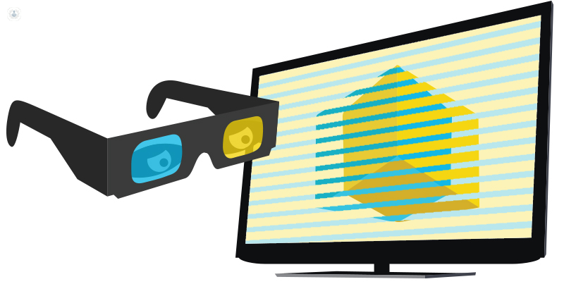
Stereoscopy or stereoscopic imaging is a technique used to collect three-dimensional (3D) images and create the illusion of depth through the stereographic image or stereogram. Two slightly offset images are shown separately to each eye of the viewer. Both images are the same but from a slightly different angle or perspective. The brain is then tricked into thinking that the two flat images shown together are one image in 3D. Special eyewear (polarized glasses) or liquid crystal shutter glasses are needed for the brain to make sense of the picture.
What does it consist of?
There are three ways that stereoscopy can be done.
- Two images can be shown separately and shutter glasses used to filter the image so that the correct eye sees it.
- Both images are superimposed on each other and polarized glasses are used to combine the images.
- Each image is shown directly into each eye, without the need for glasses, done using a parallax barrier on a screen, allowing both eyes to see different images.
Why is it done?
Stereoscopy can be applied to several fields. In the case of medicine, it helps to visualize the width, length and depth of images of the interior of the body from images obtained by CT or Nuclear Magnetic Resonance, both for teaching or research and for diagnosis and intervention.
Stereoscopy helps to reliably detect foreign bodies and abnormalities, expanding the information that a traditional radiograph would provide. Stereoscopes are used by optometrists, ophthalmologists and visual therapists to diagnose and treat binocular vision and visual disorders.
Meaning of abnormal results
If abnormalities are found after stereoscopy, your doctor will go over the results with you and advise you on the course of treatment needed for your particular case.