DEXA scan

Professor Gordana Prelevic - Endocrinology, diabetes & metabolism
Created on: 09-18-2018
Updated on: 04-11-2023
Edited by: Aoife Maguire
What is a DEXA scan?
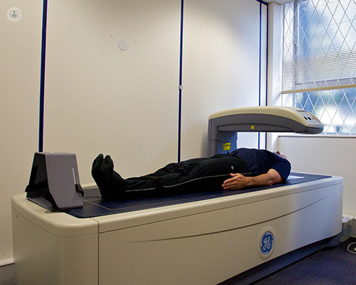
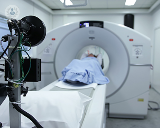
A dual energy X-ray absorptiometry (DEXA) scan, (or bone density scan) is a technique used to measure bone density. The scan is quick and painless and involves the patient lying on their back on an X-ray table. There is no tunnel to pass through as with an MRI or CT scan, so there is no feeling of claustrophobia.
What is the procedure for a DEXA scan?
The scan takes around 15 minutes. Whilst lying down on an X-ray table, a scan will be carried out by a radiographer. A large scanning arm passes over the body to measure bone density in the centre of the skeleton. A low-dose beam of X-rays passes through the part of the body being examined, which is usually the lumbar spine and hip.
Some of the X-rays that pass through the body are absorbed by the bone and soft tissue. An X-ray detector inside the scanning arm is able to measure the amount of X-rays that have passed through the body, which then produces an image of the scanned part of the body that is sent to a computer.
What do my DEXA scan results show?
The bone mineral density measurements will be compared with the bone density of a young healthy adult (known as a T score) or with an adult of your age, gender and ethnicity (known as a Z score).
The comparison of the patients BMD to bone density of a healthy young adult is expressed as a T-score (represents number of standard deviations from mean young adult BMD).
The T- score of:
Above -1.0 is normal
- Between -1.0 and -2.5 defines reduced bone mineral density compared to young adults
- At or below -2.5 defines osteoporosis
Z-score represents number of standard deviations from the mean bone density of the persons of the same age. If a Z- score is below -2.0 then the bone density is lower than expected for age.
Who should have a DEXA scan?
A DEXA scan is advised for those who have a bone fracture following a minor injury. It is also advised if a patient is considered at increased risk of osteoporosis.
Recognized risks for osteoporosis are: Oestrogen deficiency in women (menopause or a lack of periods in young women), hypogonaidsm (low testosteorne level) in men, steroid treatment, smoking, excessive alcohol intake, low body weight (BMI below 19), a family hisotry of osteoporosis, a parental history of hip fracture, low calcium intake, vitamin D deficiency etc.
If the doctor thinks a patient is at risk of developing osteoporosis they may use the Fracture Risk Assessment Tool (FRAX). FRAX gives predictive likelihood of having a bone fracture following minor trauma (so called fragility fracture) within the next 10 years. This calculation tool has important role in making decision about the management of individual patients.
The DEXA scan is also used to monitor if a current treatment plan for osteoporosis is working.

Insights into DEXA scans: Assessing bone health simplified
By Dr Taher Mahmud
2024-04-27
In his latest online article, Dr Taher Mahmud gives us his insights into DEXA scan. A DEXA scan, known as dual energy X-ray absorptiometry or DXA, is an X-ray procedure specifically designed to assess bone density. This imaging technique is commonly referred to as a bone density scan or bone densitometry scan. See more
Experts in DEXA scan
-

Professor Waqar Bhatti
RadiologyExpert in:
- Sports Reviews
- MRI
- DEXA scan
- Spine MRI
- Musculoskeletal ultrasound
- Sports injuries
-

Professor Richard Keen
RheumatologyExpert in:
- Osteoporosis
- DEXA scan
- Injections for osteoporosis
- Medicolegal
- Osteoporosis diet
- Hyperparathyroidism
- See all

The Alexandra Hospital - part of Circle Health Group
The Alexandra Hospital - part of Circle Health Group
Mill Ln, Cheadle
No existe teléfono en el centro.
By using the telephone number provided by TOP DOCTORS, you automatically agree to let us use your phone number for statistical and commercial purposes. For further information, read our Privacy Policy
Top Doctors
The Highfield Hospital - part of Circle Health Group
The Highfield Hospital - part of Circle Health Group
Manchester Road, Rochdale, Lancashire, OL11 4LZ
No existe teléfono en el centro.
By using the telephone number provided by TOP DOCTORS, you automatically agree to let us use your phone number for statistical and commercial purposes. For further information, read our Privacy Policy
Top Doctors
Alliance Medical Ltd Manchester Airport
Alliance Medical Ltd Manchester Airport
Kingsley Hall, 20 Bailey Lane, Wythenshawe. M90 4AN
No existe teléfono en el centro.
By using the telephone number provided by TOP DOCTORS, you automatically agree to let us use your phone number for statistical and commercial purposes. For further information, read our Privacy Policy
Top Doctors
-
The Alexandra Hospital - part of Circle Health Group
Mill Ln, Cheadle, CheadleExpert in:
- Hip
- Cardiology
- Shoulder and elbow
- Paediatrics
- Foot and ankle
- Knee
-
The Highfield Hospital - part of Circle Health Group
Manchester Road, Rochdale, Lancashire, OL11 4LZ , RochdaleExpert in:
- Cardiology
- Orthopaedic surgery
- Plastic surgery, reconstructive and aesthetics
- Dermatology
- Diagnostic Imaging
- Endocrinology
-
Alliance Medical Ltd Manchester Airport
Kingsley Hall, 20 Bailey Lane, Wythenshawe. M90 4AN, ManchesterExpert in:
- Ultrasound
- Musculoskeletal ultrasound
- Musculoskeletal imaging
- Brain MRI
- Spine MRI
- See all
- Most viewed diseases, medical tests, and treatments
- Perimenopause
- Premature menopause
- Thumb joint replacement
- Thumb arthritis
- Periacetabular osteotomy (PAO)
- Digestive diseases
- Stress
- Women's health
- Cephalometric
- Robotic surgery