A guide to different types of knee injuries
Escrito por:Knee injuries can occur commonly, particularly when playing sport. Renowned consultant trauma and orthopaedic surgeon Mr Cristian Nita provides a guide to the most commonly ocurring knee injuries.
How serious is a knee cartilage injury?
Intra-articular cartilage lesions are very serious.
These can be divided into:
Meniscus injuries
Articular cartilage injuries
They can also be further divided into:
- trauma related: i.e. sport injuries, road traffic accident etc.
- spontaneous, non-traumatic injuries
- degenerative, age-related injuries
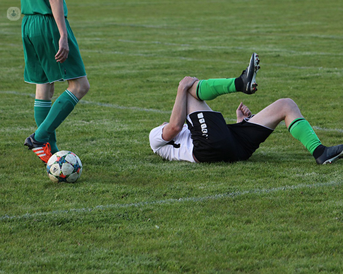
Trauma related injuries can occur following an abnormal movement of the articular surfaces on to each other. Injury is often seen following a valgus stress or pivot shift mechanism, when the foot is planted on the ground, the knee is pushed inwards, and the body momentum produces a rotational movement at the knee joint level. This forces the articular surfaces against each other leading to significant stress onto meniscus, ligaments and cartilage itself. This type of injury commonly occurs while playing sports such as football and rugby.
Similar injuries can occur when the player jumps up, lands on one leg and the knee joint jerks inwards due to the body weight supported by the leg. This commonly occurs in sports such as basketball and netball.
Diagnosis is established following examination and MRI scan. In certain cases, simple knee x-rays can indicate an intra-articular pathology. However, in the majority of cases simple x-rays are normal.
Spontaneous, non-traumatic cartilage lesions.
These can be grouped in:
Osteochondritis dissecans
These injuries can occur at all ages and most categories of people. However, teens, young adults and middle-aged patients are at higher risk of suffering from these injuries.
These injuries can be classed as:
- cartilage only, where the articular cartilage detaches from the bone and creates a hole in the articular surface
- cartilage and bine, when a fragment of bone with articular cartilage becomes detached and floats freely into the joint.
Both types can cause locking, the join may be stuck in flexed(bent) or extended position.
A diagnosis is made predominantly by MRI. Sometimes, an x-ray may indicate pathology if a bony fragment is involved.
Spontaneous osteonecrosis
These usually occur suddenly, with significant pain, swelling and inability to weight bear painlessly.
Pre-existing conditions which require corticosteroid intake are known to contribute to the incidence.
Certain professions such as divers, are known to be at a higher risk of developing spontaneous osteonecrosis.
To diagnose spontaneous osteonecrosis in the early stages an MRI is taken. In later stages, an x-ray is taken.
Pathophysiologic mechanism of this is unknown and the incidence is fairly low.
The onset of symptoms is sudden and associated with pain, swelling, locking or pseudo-locking. Minor trauma such as a sprain can initiate symptoms.
Degenerative cartilage lesions
These occur naturally with age and use. The development of these may be provoked by activities such as extensive running, i.e. marathon runners, fell walking, etc.
The onset of symptoms is gradual and progressive. Symptoms include an aching pain associated with swelling and sleep disturbance.
Participating in activities such as walking, descending stairs and sitting for long will worsen symptoms. These are also frequently associated with stiffness after resting a period of time and can vary in intensity and duration, with good periods and bad periods.
Articular cartilage
The articular cartilage covers the ends of the bone. It protects the bones and permits smooth joint movement.
If trauma occurs, the articular cartilage can break away which in turn, blocks the normal range of movement and locks the knee. It can also wear away and the joint begins to swell, causing pain. The patient’s movement will then become stiff, leading to reduce mobility.
Meniscus
Injuries to the menisci may occur due to certain movements in the knee. These injuries can occur if the knee is extended very suddenly. There is no time for the meniscus to move forward, leaving it trapped between the femoral condyle and the tibial plateau.
Meniscus tears are extremely common, particularly among athletes. There are several types of tears, including longitudinal tears, parrot beak, flap, bucket handle and mixed/complex. If the injury has developed due to sport, it is often presented alongside ligament injuries.
The diagnosis is established using simple x- rays. Presence of mechanical symptoms such as catching, locking are further investigated with MRI scan and final diagnosis is established.
If you are suffering from a knee injury and would like to speak to Mr Nita, do not hesitate to book an appointment through his Top Doctors profile today.