Insights into DEXA scans: Assessing bone health simplified
Escrito por:In his latest online article, Dr Taher Mahmud gives us his insights into DEXA scan. A DEXA scan, known as dual energy X-ray absorptiometry or DXA, is an X-ray procedure specifically designed to assess bone density. This imaging technique is commonly referred to as a bone density scan or bone densitometry scan.
The purpose of a DEXA scan:
Primarily, a DEXA scan is employed to diagnose osteoporosis or evaluate the risk of its development. Osteoporosis, a condition that weakens bones, can be identified and evaluated through this scan, providing insights into bone density.
Who should undergo a DEXA scan for osteoporosis?
Individuals over 50, particularly those at risk of osteoporosis, are recommended to undergo a DEXA scan. Factors elevating the risk include a family history of fractures and, for those under 50, additional risk elements such as smoking, excessive alcohol consumption, or a history of bone fractures. Moreover, women experiencing early menopause or having undergone it without hormone replacement therapy, individuals with conditions like arthritis causing low bone density, those on medications affecting bone strength, and women with irregular menstrual cycles may also benefit from a DEXA scan.
Procedure for a DEXA scan:
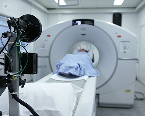
The scan is quick, painless, and generally allows patients to remain clothed, except for items containing metal, which need to be removed. No specific preparation, fasting, or dietary alterations are required. During the scan, individuals lie on a flat, open X-ray table, eliminating any sensation of confinement typically associated with MRI machines. A specialised scanner passes over the body, emitting a low-dose X-ray beam to measure bone density, typically focusing on areas like the hip, spine, and wrist. Scans are brief, taking around five minutes, with no need for extended hospital stays.
Interpreting DEXA scan results:
DEXA scan results provide a 'score' indicating bone density, relative to healthy levels adjusted for age, ethnicity, and gender. This score, known as the T score, categorises results as Normal (above -1 deviation), Slightly reduced (between -1 and -2.5 deviation), or indicative of osteoporosis (at or below -2.5 deviation). It's important to note that while these results gauge bone strength, they do not definitively predict fracture risk. Even individuals with normal bone density may experience fractures. Post-scan, your doctor analyses the results, considers individual risk factors, and then determines the necessity and course of any potential treatments or interventions.
Enhancing and simplifying the understanding of DEXA scans:
Explaining the purpose, procedure, and interpretation of DEXA scans in simpler terms can greatly benefit individuals seeking this evaluation. Furthermore, emphasising the role of risk factors and the necessity for a doctor's interpretation can enhance comprehension and promote informed decision-making regarding bone health.
Dr Taher Mahmud is a highly regarded rheumatologist with over 30 years of experience. You can schedule an appointment with Dr Mahmud on his Top Doctors profile.