An expert explains: Ultrasound guided surgery for endometriosis
Autore:The use of ultrasound scan imaging in treatment for endometriosis improves surgical accuracy and precision as well as offering better outcomes for patients. In this informative article, leading consultant in gynaecology and gynaecological oncology surgery Mr Ahmad Sayasneh sheds light on the applications of ultrasound imaging in gynaecological surgery and offers expert insight on how the use of this technology benefits patients suffering from endometriosis.

What is involved in image coded surgery for endometriosis?
The term ‘image coded’ relates to using imaging technology to increase the precision and accuracy of a surgical procedure. The ultrasound scan, which takes places while the operation is being carried out, allows the surgeon to better identify any disease and ensure that it is fully treated. This is particularly useful in cases of endometriosis as well as some other gynaecological conditions where the areas of disease need to be identified and excised. The use of imaging ensures that any disease can be accurately removed and that nothing is accidentally left behind.
What are the advantages of using imaging in this type of surgery?
Ultrasound imaging gives the surgeon deeper and more detailed vision so that the disease can be assessed with greater precision. For example, if there is endometriosis in the retroperitoneal space (the area behind the peritoneum), around the roots of the nerves or in spaces that aren’t visible during keyhole surgery or laparoscopy, an ultrasound scan allows the surgeon to visualise the disease.
Once visualised, which may be achieved through transrectal or transvaginal ultrasound scanning, the surgeon is able to remove any disease accurately without opening unnecessary spaces, which can increase recovery time after the procedure. The benefit for the patient, therefore, is enormous as the disease can be targeted accurately and the surgeon can ensure that nothing is left behind.
Is image coding always necessary in surgery for endometriosis?
In many cases, endometriosis surgery can be successfully performed without using intraoperative imaging. This is simply because in some cases, the disease is well visualised during the surgery itself and there is therefore no need for ultrasound imaging. However, when the surgeon cannot visualise the disease, imaging is required to ensure the operation is a success.
In some cases, surgeons are able to review images taken prior to surgery in order to map out how the procedure should take place. In more complex cases, however, ultrasound scanning during the surgical procedure is a very useful tool that allows the surgeons to work precisely to remove disease. There is no ideal candidate for image coding in surgical procedures and any patient may benefit from it, whether being treated for endometriosis or any other gynaecological pathology. It's also very useful when treating ovarian cysts to target the exact areas of disease, particularly when they are small in size and hard to visualise.
Does image coding pose any additional risk to the patient?
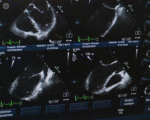
Image coding merely complements usual surgical procedures and so it is very safe and poses no additional risk to the patient. Much like any other tool used in surgery, it aids the surgeon to complete the required procedure with improved accuracy and safety. During the procedure, the ultrasound scan is shown on a screen, additional to the one usually used in keyhole surgery or laparoscopic surgery, allowing the surgeon to map the procedure precisely.
If you require treatment for a gynaecological condition, such as endometriosis, and wish to book a consultation with Mr Sayasneh to discuss your options, you can do so by visiting his Top Doctors profile.


