
What's the difference between 2D, 3D and 4D ultrasound?
Written by:What is ultrasound used for? An ultrasound scan is a diagnostic technique which has many purposes though it is typically used by a gynaecologist to monitor the foetus in the mother's womb during pregnancy.
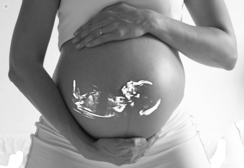
How does ultrasound work?
Sound is a transmission wave. When sound hits a surface that can reflect it, the sound (or echo) is bounced back to the source. The ultrasound machine is the source of the sound waves and also collects them to produce them as visual images on a screen.
What are the differences between 2D, 3D and 4D ultrasound?
3D and 4D ultrasounds allow imaging and three-dimensional analysis. This makes it possible for patients to recognise the images that appear on the monitor, which was not the case with 2D ultrasounds, when only the specialist was able to interpret the images. Even so, these ultrasounds involve additional training and also require an important technological investment. Other advantages of 3D and 4D ultrasound over 2D are that it is possible to study musculoskeletal malformations more closely, undertake an evaluation of facial dimorphism and also study abnormalities in hands and feet. It is possible to evaluate cranial sutures, look bone fractures and detect subtle anomalies of the vertebrae. In addition to obtaining and displaying images in the same way as 3D, 4D ultrasound captures images in real time.
During a pregnancy, you will generally have a 12 week ultrasound, although generally, they should be done between weeks 20 and 22. With the 4D ultrasound, it is possible to visualise the baby’s face much earlier than this, making an early pregnancy scan more useful. 4D ultrasound also helps with prenatal diagnosis of congenital abnormalities, although genetic diseases can only be detected by studying the karyotype (the chromosomal pattern of an individual).
Is the image resulting from a 4D ultrasound always clear?
As the foetus grows, the space in the womb and the baby's mobility decrease, which can make visualisation difficult. It also depends heavily on the foetal position, amniotic fluid, placenta and the body mass index (BMI) of the pregnant woman, since the ultrasound is not so effective on obese pregnant women. However, these ultrasounds create good contact between mother, child and father, making emotional bonds that will be enhanced after birth, benefiting child development and maturation.
4D scans represent an advance in the diagnosis of many morphological diseases and therefore it must be used by specialists who can analyse beyond the face of the foetus, using analysis of amniotic fluid, placenta and foetal-placental circulation, to perform diagnostics and biometrics.