What is PUJ obstruction?
Written by:Some children are born with a condition called pelviureteric junction (PUJ) obstruction, which blocks the flow of urine from their kidneys to the bladder. Serious blockage can lead to a deterioration of the function in that kidney , so we asked consultant paediatric urologist Miss Marie-Klaire Farrugia how PUJ obstruction is diagnosed, and when surgery is considered as a treatment option.

Pelviureteric junction (PUJ) obstruction is most commonly due to a narrowing in the part of the ureter which drains urine from the kidney, causing a build-up of urine which then increases the pressure within the kidney - with the potential to cause damage.
PUJ obstruction is most often a congenital condition, occurring in 1 in 1500 births. It tends to affect the left kidney more than the right, but in 10% of cases it affects both kidneys.
What causes PUJ obstruction?
PUJ obstruction can be:
- intrinsic - the blockage occurs inside, e.g. due to stenosis (a narrowing of the passage)
- extrinsic – the blockage is caused by an external factor putting pressure on the PUJ, such as an extra blood vessel crossing over the ureter.
How is PUJ obstruction diagnosed?
In most cases, PUJ obstruction is diagnosed before birth, because it causes a dilatation in the urinary tract, which can be seen on prenatal scans. However, it is possible to get a PUJ obstruction later in childhood or even in adulthood, in which case symptoms can include a sharp flank pain accompanied by nausea and vomiting; urinary tract infections; or swelling in the abdomen.
To make a diagnosis, a radiologist will carry out an ultrasound scan of the kidneys and bladder. If your child has a PUJ obstruction, the ultrasound usually shows a swelling, or dilatation, of the kidney, and in some cases a thinning in the outermost layer of the kidney which is being compressed by the increased pressure.
The next stage is to carry out a MAG-3 renogram, which is a more specific test to assess the function and drainage of the affected kidney.
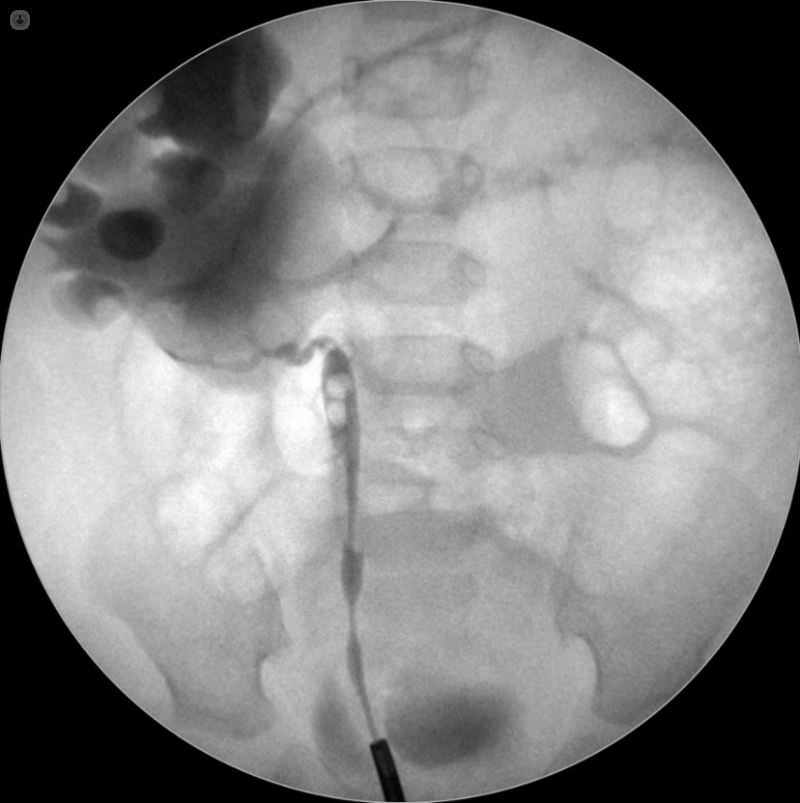
If the diagnosis is still not confirmed, other options include an MRI of the urinary tract or a retrograde pyelogram to see the urinary tract anatomy and urine drainage more clearly.

How is PUJ obstruction treated?
PUJ obstruction is not always treated in the first instance, because the obstruction may not be significant enough to cause kidney damage or symptoms. Your child will be monitored on a regular basis with ultrasound and the option of a repeat MAG-3 renogram, and in the case of a child with severe dilation, antibiotics may be prescribed.
In some cases, the obstruction can resolve on its own and this will be clear on the ultrasound scans.
Surgery is generally recommended:
- In the presence of symptoms such as pain, haematuria, or infection
- where there is a deterioration in kidney function
- where there is progression of the kidney dilatation
PUJ obstruction is treated by means of a procedure called pyeloplasty. The procedure can be performed with an open incision or via laparoscopy, with or without robotic assistance. Recovery is usually within 72 hours. Follow-up after the surgery will include a further ultrasound and MAG-3 renogram – once these confirm success of the surgery, your child will be discharged. The success rate of the procedure is over 95%.


