Image guided radiotherapy (IGRT)

What is image-guided radiotherapy (IGRT)?

Image-guided radiotherapy (IGRT) is a technique in which high-precision three-dimensional images are taken before and during radiotherapy to show the precise size, shape and location of the tumour. It is a technique that allows greater precision in the delivery of radiation to the tumour, which means a higher dose can be delivered to the cancerous cells without irradiating the surrounding healthy cells.
 

Why is image-guided radiotherapy done?

Imaging-guided radiation therapy (IGRT) is used to treat tumours located in areas of the body normally prone to movement (for example, lungs, liver, prostate, etc.) or those located near vital organs and tissues.

Notably, it is a technique that is often combined with other advanced forms of high-precision radiation therapy, such as intensity-modulated radiation therapy (IMRT), proton beam therapy, stereotactic radiotherapy or stereotactic body radiotherapy (SBRT).


What does image-guided radiotherapy consist of?

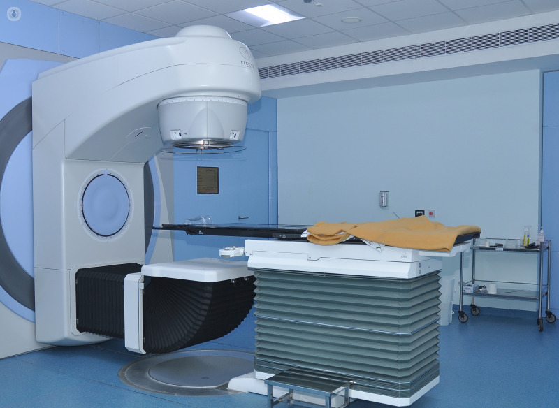
IGRT consists of taking images using X-rays, CT scans, and perhaps MRI or PET scans in order to map out the exact size, shape and location of the tumour and thus improve the precision of the treatment and preserve the surrounding healthy tissues. Scans are taken before and during the administration of the radiotherapy, while the patient is lying on the treatment table. Scanning equipment is often built into or mounted on the machine that delivers the radiotherapy, e.g. linear accelerators.

Scans taken during the procedure are referred to as 4D-RT, the fourth dimension being time. This is because the scans allow the treatment team to see if the tumour moves during the scan and follow it with the radiation beam. This makes it a useful technique for tumours on organs that move, such as the lungs.


Aftercare

Since image-guided radiation therapy allows the radiation to be delivered very precisely to the tumour, only a small area of the surrounding healthy tissue will receive any radiation, reducing the chances of side-effects. However, side-effects can still occur and the radio oncology expert will recommend periodic visits to monitor the patient’s progress.

09-11-2023
Top Doctors

Image guided radiotherapy (IGRT)

Dr Irene De Francesco - Clinical oncology

Created on: 02-08-2013

Updated on: 09-11-2023

Edited by: Conor Dunworth

What is image-guided radiotherapy (IGRT)?

Image-guided radiotherapy (IGRT) is a technique in which high-precision three-dimensional images are taken before and during radiotherapy to show the precise size, shape and location of the tumour. It is a technique that allows greater precision in the delivery of radiation to the tumour, which means a higher dose can be delivered to the cancerous cells without irradiating the surrounding healthy cells.


Why is image-guided radiotherapy done?

Imaging-guided radiation therapy (IGRT) is used to treat tumours located in areas of the body normally prone to movement (for example, lungs, liver, prostate, etc.) or those located near vital organs and tissues.

Notably, it is a technique that is often combined with other advanced forms of high-precision radiation therapy, such as intensity-modulated radiation therapy (IMRT), proton beam therapy, stereotactic radiotherapy or stereotactic body radiotherapy (SBRT).


What does image-guided radiotherapy consist of?

IGRT consists of taking images using X-rays, CT scans, and perhaps MRI or PET scans in order to map out the exact size, shape and location of the tumour and thus improve the precision of the treatment and preserve the surrounding healthy tissues. Scans are taken before and during the administration of the radiotherapy, while the patient is lying on the treatment table. Scanning equipment is often built into or mounted on the machine that delivers the radiotherapy, e.g. linear accelerators.

Scans taken during the procedure are referred to as 4D-RT, the fourth dimension being time. This is because the scans allow the treatment team to see if the tumour moves during the scan and follow it with the radiation beam. This makes it a useful technique for tumours on organs that move, such as the lungs.


Aftercare

Since image-guided radiation therapy allows the radiation to be delivered very precisely to the tumour, only a small area of the surrounding healthy tissue will receive any radiation, reducing the chances of side-effects. However, side-effects can still occur and the radio oncology expert will recommend periodic visits to monitor the patient’s progress.

This website uses our own and third-party Cookies to compile information with the aim of improving our services, to show you advertising related to your preferences as well analysing your browsing habits. You can change your settings HERE.