The role of imaging in congenital heart diseases
Escrito por:Cardiovascular imaging has a pivotal role in the lifelong management of coronary heart disease (CHD) patients. The improvements in medical imaging and surgical or percutaneous procedures have resulted in an increase in survival rates amongst these patients.
We spoke to Professor Giovanni Di Salvo to ask why medical imaging is so important in the care of congenital heart disease and how often patients need to get checked. Read on to find out more.

Why is it so important and what type of imaging is used?
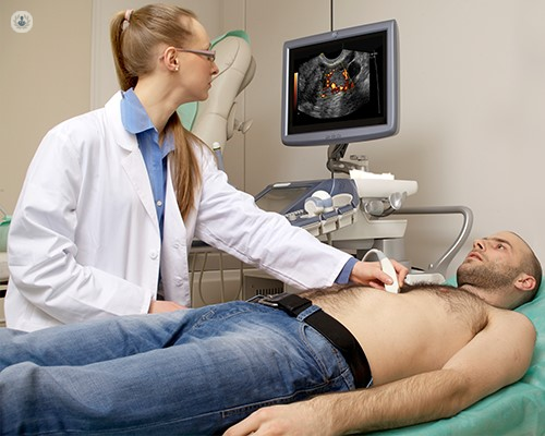
All CHDs need to undergo imaging nowadays, and it is only after performing complete and expert imaging, can we then define the right management for the patient. The first-line and central imaging modality is an echocardiogram. This technique is safe, radiation-free, repeatable and provides us with a lot of invaluable information. If we see from the echocardiography that additional imaging is needed, then a cardiac MRI or CT scan can also be performed.
Only an expert in diagnosing and treating adult congenital heart disease can decide which imaging pathway is needed for that particular patient and which particular information that imaging technique needs to provide.
In paediatric cases, echocardiography is usually the only imaging modality we need to perform; however, in adults with CHD, echocardiography is very often integrated with cardiac magnetic resonance imaging (CMRI). Generally, the more complex the congenital heart disease is, the more likely it is that will require a multimodality imaging approach.
What is imaging used for?
Imaging can be used for various reasons to create detailed pictures of inside your body to examine your heart and blood vessels. Therefore, imaging can help a doctor to:
- Diagnose many different heart conditions
- Help plan any subsequent surgical or percutaneous procedures
- Assess how well the heart is working
- Evaluate residual lesions
- Evaluate the success of any surgical repair
How often should people with congenital heart diseases get a check-up?
This depends entirely on the patient and their specific problem. Generally, though, patients should undergo a scan or a check-up every six months particularly for more complex diseases. This can be reduced to annually for less complex lesions or stable patients. If their clinical scenario changes, then a closer follow up is typically needed.
What advances are there in non-invasive imaging for congenital heart disease?
There have been many advances in non-invasive imaging, from live 3D echocardiography to speckle tracking echocardiography - which analyses the motion of tissues in the heart and provide information which is not available from traditional echocardiography.
Cardiac MRI scans can provide exceptional levels of detail of the anatomy and extracardiac and tissue characterisation which has important implications for the patient’s outcome.
Cardiac CT scans are crucial to assess coronaries and small vessels as well as in patients with a metallic prosthesis or stent when CMRI is less of a value. Additionally, CT scans are more comfortable for patients who suffer from claustrophobia or the fear of enclosed spaces, as the machines are shorter and less noisy than that of a cardiac MRI scan.
To book an appointment with Professor Giovanni Di Salvo, visit his Top Doctors profile and check his availability.


