What is transcranial magnetic stimulation (TMS)?
Escrito por:In his last article for Top Doctors, Professor Keyoumars Ashkan, leading neurosurgeon at the London Neurosurgery Partnership, introduced us to some of the latest developments in the surgical treatment of brain tumours. One such development is TMS, or transcranial magnetic stimulation, which is used across several medical specialties and is proving effective as a tool in the surgical removal of brain tumours. Professor Ashkan talks us through how TMS works, and what it is used for.
What is TMS?
Transcranial magnetic stimulation, otherwise known as TMS, is a procedure which uses a magnetic field generator to stimulate particular areas of the brain. This coil produces small electric currents in regions of the brain, and it is a non-invasive method of brain stimulation which is proving useful in the management of various conditions and pathologies.
It can be used diagnostically to measure brain activity and function, and evaluate damage such as from stroke, multiple sclerosis, motor neuron disease, and other disorders which affect the spinal cord and facial/cranial nerves.
In terms of treatment, rTMS (repetitive TMS) is proving useful in treating neuropathic pain, which currently has few effective treatments available, and it is now being trialled as an alternative or adjunct to drugs to treat depression in certain patients.
How can TMS help in the treatment of brain tumours?
In neurosurgery, navigated transcranial magnetic stimulation has proven useful in mapping tumours in the brain, checking the exact relationship between the tumour and particular areas of the brain. Certain regions control different things, such as parts responsible for movement, and parts responsible for speech. When planning the removal of a tumour, it is essential for the neurosurgeon to understand where the tumour sits in relation to these critical areas in the brain.
Mapping the brain in this way allows for the accurate removal of brain tumours without affecting functional areas. It allows visualisation the brain, drawing a map of the nerves and effectively guiding the surgeon to where they are able to operate, and where they must avoid. This also allows for more precise surgery, with smaller incisions.
How is TMS performed?
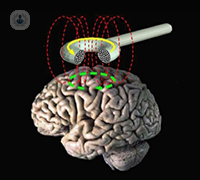 In order to be able to define motor areas in the brain, a magnetic coil is used to scan specific locations. It is passed over these locations in the head, inducing short sharp electrical pulses, which stimulate the neurons in those particular areas. These pulses then activate motor neurons in the brain, which trigger parts of the body into reacting and allow for muscle activity to be measured, using electrodes placed on the arms and legs.
If muscle activity is measured, the areas scanned are then flagged as points to avoid as they indicate motor connection. Similar techniques can also be used to map the speech and language areas in the brain. This brain mapping technique is especially useful, as it gives a clear idea of the individual’s brain – tumours can often displace certain areas, meaning it is essential for the neurosurgeon to have a clear idea of how the brain is mapped out.
The procedure is non-invasive and therefore causes minimal discomfort, if at all.