MRI

What is an MRI?

MRI (magnetic resonance imaging) is a type of scan that doctors use to see the bones, tissues, and organs inside your body. MRI uses strong magnetic fields and radio waves to create highly detailed images, much more detailed than a CT scan or X-ray. This can be very useful when making a diagnosis, or planning surgery.

Some of the most common types of MRI scan include:

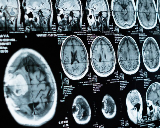
  • Brain MRI – often used to assess damage to the brain following injury or a stroke, detect tumours, or diagnose degenerative diseases such as Alzheimer’s disease.
  • Spinal MRI – used to diagnose degenerative problems in the spine or identify slipped disk, fractures, inflammation, or tumours.
  • Abdominal MRI – most often used to diagnose kidney problems but also useful for detecting issues with the liver or gall bladder.
  • Breast MRI – usually used in women who have been diagnosed with breast cancer to determine the size and position of the tumour, or tumours.
What does an MRI involve?

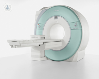
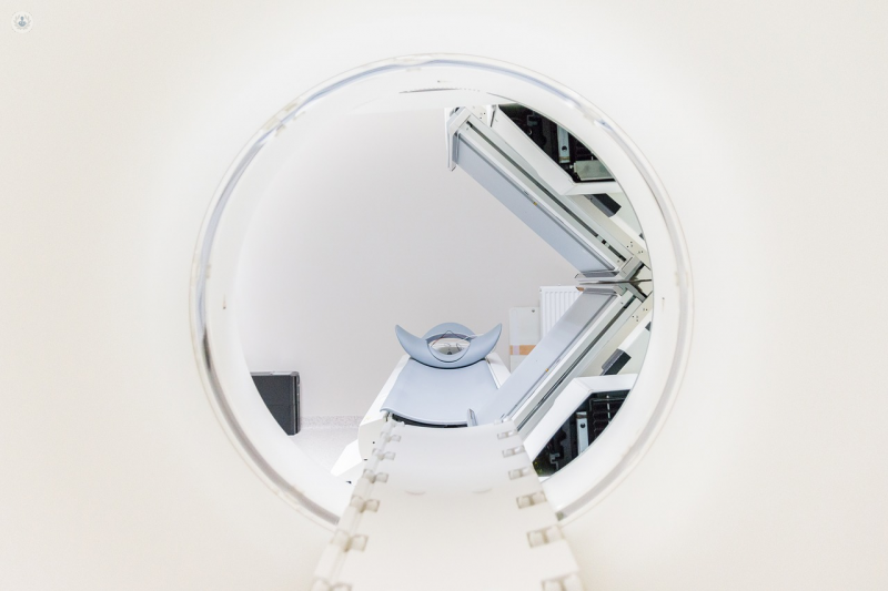
An MRI scanner is a large tube surrounded by magnets. The scanner can be used to produce images of any part of your body. An MRI scan involves lying down on a flat bed inside the scanner for a period of time, usually between 15 and 90 minutes, while a radiologist takes several pictures of your body. Each picture can take a few minutes, so it’s important to be as still as possible during the scan.

Preparing for an MRI

Even though an MRI is one of the safest procedures available, not everyone can have an MRI. Because the MRI scanner generates a strong magnetic field, it can be risky if you have something metallic in your body. See this guide from the NHS for a full list of metal implants to consider.

It’s equally important to remove anything metal that you’re wearing, so any jewellery, piercings, hairpins or clothes with metal fasteners should be left at home. You might be asked to wear a hospital gown for the scan.

You should tell the radiologist if you might be pregnant, since the effects of an MRI on a unborn child aren’t yet known.

If you suffer from claustrophobia you can ask for a prescription of a mild sedative to take before the scan. You’ll be transferred to a recovery room after the scan to come round.

Finally, you might receive an injection of a contrast dye to highlight certain parts of the body on the scan. This will be administered using an IV line, inserted into a vein in your hand or arm. The dye is usually safe, but might not be recommended if you have a kidney problem, or recently had surgery, so it’s important to let the doctor know if this applies to you.

What to expect during the MRI scan

Once you’ve removed any metal accessories, and you’ve received the contrast day, you’ll be positioned on the bed and moved into the scanner. You might go in feet first or head first depending on what is being scanned.

The radiologist will operate the scanner from another room, where the magnetic field cannot interfere with their computer, but you’ll be able to speak to them via a microphone placed inside the scanner. At certain points, you might be asked to hold your breath, but it will never be for more than a few seconds.

Sometimes the scanner will make a loud clicking sound as it sends out radio waves. You’ll be given headphones to wear for the noise, and you can listen to music throughout the scan.

The whole process should take between 15 and 90 minutes, depending on the size of the area being scanned and whether or not you need a contrast dye. You can leave straight after the scan, or as soon as you’ve recovered from the sedative.

What happens after the MRI scan?

What happens next will depend on your condition and the type of MRI being performed. It is unlikely you will receive your test result immediately because scan will need to be studied by the radiologist and other specialists.

02-23-2024
Top Doctors

MRI

Dr Graham Cole - Cardiology

Created on: 06-20-2013

Updated on: 02-23-2024

Edited by: Jay Staniland

What is an MRI?

MRI (magnetic resonance imaging) is a type of scan that doctors use to see the bones, tissues, and organs inside your body. MRI uses strong magnetic fields and radio waves to create highly detailed images, much more detailed than a CT scan or X-ray. This can be very useful when making a diagnosis, or planning surgery.

Some of the most common types of MRI scan include:

  • Brain MRI – often used to assess damage to the brain following injury or a stroke, detect tumours, or diagnose degenerative diseases such as Alzheimer’s disease.
  • Spinal MRI – used to diagnose degenerative problems in the spine or identify slipped disk, fractures, inflammation, or tumours.
  • Abdominal MRI – most often used to diagnose kidney problems but also useful for detecting issues with the liver or gall bladder.
  • Breast MRI – usually used in women who have been diagnosed with breast cancer to determine the size and position of the tumour, or tumours.

What does an MRI involve?

An MRI scanner is a large tube surrounded by magnets. The scanner can be used to produce images of any part of your body. An MRI scan involves lying down on a flat bed inside the scanner for a period of time, usually between 15 and 90 minutes, while a radiologist takes several pictures of your body. Each picture can take a few minutes, so it’s important to be as still as possible during the scan.

Preparing for an MRI

Even though an MRI is one of the safest procedures available, not everyone can have an MRI. Because the MRI scanner generates a strong magnetic field, it can be risky if you have something metallic in your body. See this guide from the NHS for a full list of metal implants to consider.

It’s equally important to remove anything metal that you’re wearing, so any jewellery, piercings, hairpins or clothes with metal fasteners should be left at home. You might be asked to wear a hospital gown for the scan.

You should tell the radiologist if you might be pregnant, since the effects of an MRI on a unborn child aren’t yet known.

If you suffer from claustrophobia you can ask for a prescription of a mild sedative to take before the scan. You’ll be transferred to a recovery room after the scan to come round.

Finally, you might receive an injection of a contrast dye to highlight certain parts of the body on the scan. This will be administered using an IV line, inserted into a vein in your hand or arm. The dye is usually safe, but might not be recommended if you have a kidney problem, or recently had surgery, so it’s important to let the doctor know if this applies to you.

What to expect during the MRI scan

Once you’ve removed any metal accessories, and you’ve received the contrast day, you’ll be positioned on the bed and moved into the scanner. You might go in feet first or head first depending on what is being scanned.

The radiologist will operate the scanner from another room, where the magnetic field cannot interfere with their computer, but you’ll be able to speak to them via a microphone placed inside the scanner. At certain points, you might be asked to hold your breath, but it will never be for more than a few seconds.

Sometimes the scanner will make a loud clicking sound as it sends out radio waves. You’ll be given headphones to wear for the noise, and you can listen to music throughout the scan.

The whole process should take between 15 and 90 minutes, depending on the size of the area being scanned and whether or not you need a contrast dye. You can leave straight after the scan, or as soon as you’ve recovered from the sedative.

What happens after the MRI scan?

What happens next will depend on your condition and the type of MRI being performed. It is unlikely you will receive your test result immediately because scan will need to be studied by the radiologist and other specialists.

This website uses our own and third-party Cookies to compile information with the aim of improving our services, to show you advertising related to your preferences as well analysing your browsing habits. You can change your settings HERE.