Brain MRI

Ms Anna Miserocchi - Neurosurgery
Created on: 07-23-2015
Updated on: 11-22-2023
Edited by: Karolyn Judge
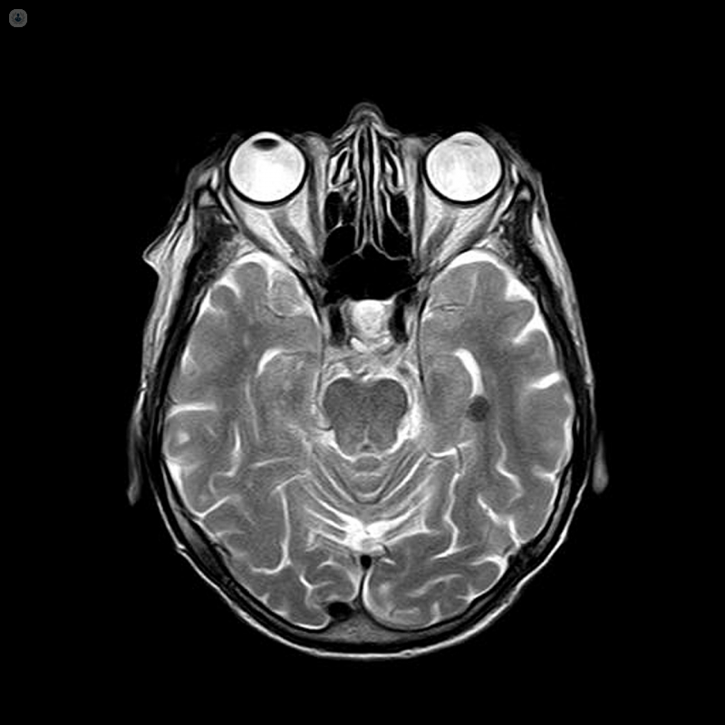
A brain MRI is a type of scan which uses a magnetic field and radio waves to produce detailed images of your brain. An MRI is carried out by a radiologist and is a safe and pain-free procedure.
Why do I need a brain MRI?
A brain MRI is used to investigate a number of things. If you have gone to the doctor with symptoms such as dizziness, headaches, seizures, or changes in your behaviour, a brain MRI can help to check if there are problems with the tissues in your brain. A brain MRI is also often used to determine if there is any damage to the brain after an injury or stoke, and if so, what kind of damage.
The images produced from the scan can help to diagnose the following conditions:
- aneurysms
- stroke
- haemorrhage
- multiple sclerosis
- tumours
- spinal cord injuries
- infections
- brain trauma
If the MRI machine is used to examine the blood vessels, the test is instead known as a Magnetic Resonance Angiography (MRA). However, the test still involves exactly the same process.
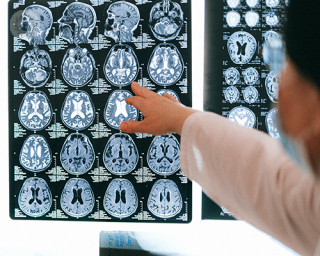
You might need to have further scans or tests after the brain MRI before a formal diagnosis can be given.
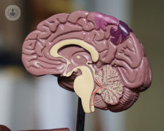
What are functional MRI scans?
A functional brain MRI (fMRI) is a special kind of brain MRI used to diagnose dementia, assess damage from a head injury, or prepare for brain surgery. The fMRI involves scanning different parts of the brain to determine which part performs which functions, in particular important communication and movement functions. To determine this you will be asked to perform basic tasks, such as answering questions, and the scan will detect what is happening in your brain while you carry these tasks out.
What can I expect from a brain MRI?
For a detailed look at how MRI tests work, what you can expect, and how to prepare, see our page on MRI scans.
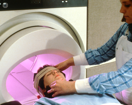
In a brain MRI, the test generally takes between 30 and 60 minutes. You might have a plastic coil placed around your head, and you might be administered a special called gadolinium through an IV. This can help doctors understand the images from the scan by highlighting different areas of the brain, particularly the blood vessels. The dye is usually safe but there may be a risk if you have problems with your kidney. You should make your doctor aware of any pre-existing conditions before they give you the IV.

White matter lesions explained by a neurologist
By Professor Hedley Emsley
2024-07-27
Professor Hedley Emsley explains what white matter lesions are, the range of potential causes, if they can cause problems and more. See more

Mild traumatic brain injuries: A modern perspective on treatment
By Dr Priyanka Pradhan
2024-07-25
We learn more about mild traumatic brain injuries and how they are treated from highly respected chartered clinical psychologist and registered clinical neuropsychologist Dr Priyanka Pradhan in this informative article. See more


Getting lost easily could be an early sign of Alzheimer’s - a expert neurologist reveals
By Professor Dennis Chan
2024-07-25
A poorer sense of direction or difficulty finding your way around places may be among the first indications that Alzheimer’s disease could affect you later on in life. These are the findings of an ongoing study conducted by experts at Edinburgh University. Consultant neurologist, Dr Dennis Chan explains more. See more
Experts in Brain MRI
-

Professor James Teo
NeurologyExpert in:
- Movement disorders
- Parkinson's disease
- Stroke
- Traumatic brain injury
- Brain MRI
- Neurorehabilitation
- See all

Cleveland Clinic London Rehabilitation Unit
Cleveland Clinic London Rehabilitation Unit
33 Grosvenor Place
No existe teléfono en el centro.
By using the telephone number provided by TOP DOCTORS, you automatically agree to let us use your phone number for statistical and commercial purposes. For further information, read our Privacy Policy
Top Doctors
-

Cleveland Clinic London Rehabilitation Unit
33 Grosvenor Place, Central LondonExpert in:
- Musculoskeletal pain
- Physiotherapy
- Neurorehabilitation
- Polytrauma
- Rehabilitation
- Specialist rehabilitation
- Most viewed diseases, medical tests, and treatments
- DEXA scan
- Ulnar nerve surgery
- Carpal tunnel surgery
- Carpal tunnel syndrome
- Peripheral nerve block
- Minimally invasive spinal surgery
- Back pain
- Spinal surgery
- Stress
- ADHD