Spine MRI

Professor Waqar Bhatti - Radiology
Created on: 07-23-2015
Updated on: 04-20-2023
Edited by: Conor Lynch
What is a spine MRI?
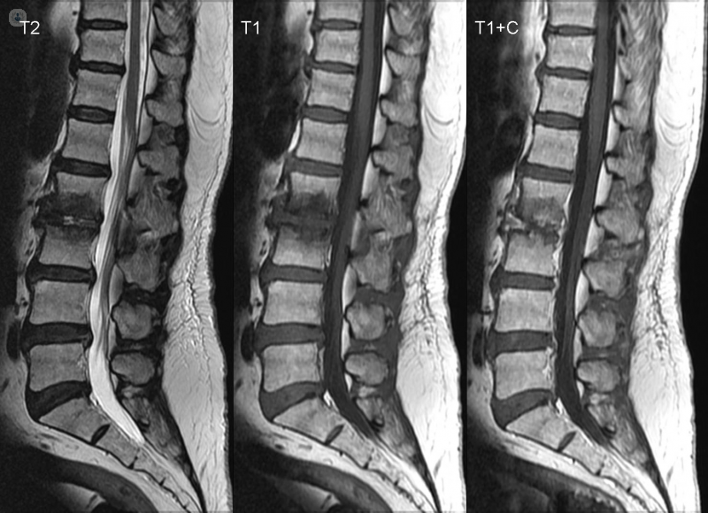
A spinal MRI is a type of scan which uses a magnetic field and radio waves to produce detailed images of your spine. An MRI is carried out by a radiologist, and is a safe procedure.
Why might I need a spinal MRI?
A spinal MRI is often advised if you suffer from back pain that won’t go away, or numbness in your legs or arms. Compared to a CT scan or X-ray, an MRI can produce a much more detailed image of your spine, giving a clear picture of the vertebrae that make up the spine, the spinal cord, discs and ligaments. A spinal MRI is therefore useful for investigating:
- any tumours present in the spine
- bone, disc or spinal cord injury
- inflammation of the spinal cord or nerves
- infection in the spine
- spinal compression or degeneration
The images produced from the scan can help to diagnose:
- slipped disc
- arthritis
- osteoporosis
- metastatic spinal cancer
However, in many cases, the doctor might need to conduct further tests to make a formal diagnosis. For example, if a tumour on the spine is found, it may not be possible to identify it as benign or cancerous with an MRI alone, meaning a further test such as a biopsy would be required. If you already have a diagnosed condition or the doctor is already aware of a tumour present in your spine, an MRI is often used to help plan for surgery.
How is a spinal MRI performed?
For a detailed look at how MRI tests work, what you can expect, and how to prepare, see our page on MRI scans. In a spinal MRI, the test can take between 30 and 60 minutes. The length of time depends on how much of your spine is scanned and whether a dye is used to highlight certain parts of the spine.
If a dye is used, this will be administered through an IV. This can help doctors understand the images from the scan by highlighting different areas of the spine. The dye is usually safe but there may be a risk if you have problems with your kidneys. You should make your doctor aware of any pre-existing conditions before they give you the IV. Finally, because it is important that the image is as clear as possible, you might need to wear straps to help you stay still during the scan.
Are there any risks associated with spine MRI scans?
There is a mimimal risk of damage to the kidneys, and as mentioned above, patients will need to let their doctor know if they have any pre-existing kidney conditions.
Experts in Spine MRI
-
Professor Waqar Bhatti
RadiologyExpert in:
- Sports Reviews
- MRI
- DEXA scan
- Spine MRI
- Musculoskeletal ultrasound
- Sports injuries
-

Dr Kathrin Hammer
RadiologyExpert in:
- MRI
- Ultrasound
- Spine MRI
- Sports injuries
- CT scan (CAT)
- Musculoskeletal ultrasound
-

Dr Katherine Miszkiel
RadiologyExpert in:
- Spine MRI
- Head CT scan
- MRI
- CT scan (CAT)
- Neuroradiology
- Brain tumour
-

Dr Simon Blease
RadiologyExpert in:
- MRI
- Ultrasound
- Spine MRI
- Musculoskeletal ultrasound
- Sports injuries
- CT scan (CAT)
-

Dr Ajay Arora
RadiologyExpert in:
- Musculoskeletal ultrasound
- CT scan (CAT)
- MRI
- Prostate MRI
- Ultrasound
- Spine MRI
- See all

Alliance Medical Harley Street
Alliance Medical Harley Street
68 Harley Street, W1G 7HE
No existe teléfono en el centro.
By using the telephone number provided by TOP DOCTORS, you automatically agree to let us use your phone number for statistical and commercial purposes. For further information, read our Privacy Policy
Top Doctors
Alliance Medical Ltd Manchester Airport
Alliance Medical Ltd Manchester Airport
Kingsley Hall, 20 Bailey Lane, Wythenshawe. M90 4AN
No existe teléfono en el centro.
By using the telephone number provided by TOP DOCTORS, you automatically agree to let us use your phone number for statistical and commercial purposes. For further information, read our Privacy Policy
Top Doctors
The Wilmslow Hospital - part of HCA Healthcare
The Wilmslow Hospital - part of HCA Healthcare
52-54 Alderley Road, Wilmslow, Cheshire
No existe teléfono en el centro.
By using the telephone number provided by TOP DOCTORS, you automatically agree to let us use your phone number for statistical and commercial purposes. For further information, read our Privacy Policy
Top Doctors
-
Alliance Medical Harley Street
68 Harley Street, W1G 7HE, W1G Marylebone LondonExpert in:
- Diagnostic Imaging
- Diagnostics
- Ultrasound
- Magnetic resonance
- Coronary calcium scan
- Coronary CT
-

Alliance Medical Ltd Manchester Airport
Kingsley Hall, 20 Bailey Lane, Wythenshawe. M90 4AN, ManchesterExpert in:
- Ultrasound
- Musculoskeletal ultrasound
- Musculoskeletal imaging
- Brain MRI
- Spine MRI
-

The Wilmslow Hospital - part of HCA Healthcare
52-54 Alderley Road, Wilmslow, Cheshire, WilmslowExpert in:
- Breast Cancer
- Men's health check
- Orthopaedic surgery
- Obstetrics and Gynaecology
- Women’s health
- Sports Medicine
- Most viewed diseases, medical tests, and treatments
- DEXA scan
- Back pain
- Medicolegal
- Cephalometric
- MRI
- OPG X-ray (Orthopantomography)
- CT scan (CAT)
- Sports injuries
- Proton beam therapy
- Port-a-cath insertion