Echocardiogram: a powerful diagnostic tool for the heart
Written by:When your heart is undergoing stress and needs to be evaluated for cardiac conditions, you may be referred to have an echocardiogram. With this procedure, your cardiologist can get a visualisation of your heart in a safe and painless way. Read on to learn about the capabilities and types of echocardiograms, as well as what the results could mean for you, as explained by Dr Malcom Burgess, a leading UK cardiologist.
When and why are echocardiograms used?
An echocardiogram is an ultrasound technique used to gather a wealth of information about the structure and function of the heart. It is relevant to many types of heart disease and one of the most frequently performed tests for patients with known or suspected heart disease.
Common reasons to perform an echocardiogram would be if there is a heart murmur detected after a physical examination or if a patient is suspected of having heart failure. Echocardiograms are also performed after a heart attack to assess the damage to the heart. Echocardiography is suited to performing follow-ups of patients with long-term cardiac conditions because it is very safe (no radiation exposure is involved), easily repeated and painless.
Are there types of echocardiogram?
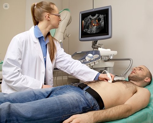
A transthoracic echocardiogram
This is the most commonly performed type of echocardiogram. It is non-invasive with the ultrasound probe being placed on the chest. Ultrasound gel is used to get good contact with the skin and to improve the image quality.
Transoesophageal echocardiogram
This is a more invasive technique reserved for selected patients. A specialised probe is passed through the mouth into the oesophagus and the ultrasound emerges from the tip. This technique is used when information from a transthoracic echocardiogram doesn’t provide enough information.
Stress echocardiogram
Sometimes, echocardiography while a patient is in a resting state only provides part of the information required. In these situations, echocardiography is performed under conditions of stress. This can be with physical exercise (using a bicycle) or using an intravenous drug to increase the patient’s heart rate. This type of test is commonly done if coronary artery disease is suspected, although coronary angiography can be provided as an alternative (using CT imaging or with an invasive catheter).
How reliable are the results?
Echocardiography provides direct visualisation of the heart on a monitor screen and it’s an accurate test. Measurements of the heart chamber sizes, wall thickness, heart function and blood flow across the valves can be made. However, the ability to make all these measurements does depend on the quality of the images and some patients are challenging to scan, meaning that sometimes limited information is available.
What is usually the next step after an echocardiogram?
Echocardiography often makes a diagnosis and depending on what is found, treatment might be required. For many patients, the test will identify a problem that does not require immediate treatment but will require follow-up scans over time to look for its progression. Occasionally, the echocardiogram might require more in-depth imaging with other techniques such as cardiac CT (computed tomography) scan or an MRI (magnetic resonance imaging) scan.
Dr Burgess is a leading cardiologist with a specialist interest in echocardiograms. Click here to see his profile and learn how he can help you improve your heart’s health.