How to prepare for a Coronary CT
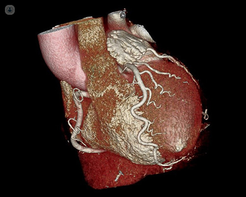
Written by:Modern CT scanners can take very high-quality pictures of the heart. The latest scanners are so fast they can take a picture of the whole heart in a less than a heartbeat. CT coronary angiography is a technique which uses CT to take pictures of the heart’s coronary arteries. Highly respected consultant cardiologist Dr Allan Harkness has been using CT for over a decade and gives lectures on the subject at national training courses. Here, he explains why it has become the go-to test NICE recommends for patients with chest pain.
Why have a Coronary CT?
Chest pain is one of the most common reasons that patients are referred to a cardiologist. The main concern both the patient and doctor have is deciding whether the pain is due to angina. Although the heart is full of blood, it needs its own separate blood supply to provide fuel and oxygen to pump efficiently.
Angina is a discomfort in the chest caused by the heart not getting enough blood to do its job. It usually comes on when you ask your heart to work harder than normal – either during exercise or if you are stressed. Classically angina is relieved quickly when you rest. If you have a GTN spray, this should take the discomfort away within a few minutes.
The typical discomfort of angina is a constricting pressure or tightness in the centre of the chest which can travel up the neck to the jaw or else down either arm – it does not have to be the left arm, although this is more common. If you have symptoms that fit this picture, then you have “typical symptoms”. If they don’t quite fit, then we say you have “atypical symptoms”.
You are more likely to have angina if your symptoms are typical than atypical. Angina is also more common in older people and in men. So, older men with atypical symptoms are actually much more likely to have angina that younger women with typical symptoms. These are:
- Smoking
- Diabetes
- High blood pressure
- High cholesterol
- A close family member with coronary artery disease
Once your cardiologist has assessed how typical your symptoms are and has considered them in the context of your age and risk factors, they will have a fair idea of how likely it is that you have angina.
In 2010, NICE only recommended coronary CT if the chances of coronary disease were low. At that time, the test was excellent at ruling out disease, but when older scanners found diseased arteries, it was sometimes difficult to decide how bad the disease was. The technology inside CT scanners has improved so much in the last few years that NICE now recommends it as the first-line test for deciding if atypical or typical symptoms are due to angina, even if the chances of significant coronary disease are high.
What preparation is needed?
When your cardiologist decided you needed a coronary CT, they would know what your heart rate is and can decide if you need any extra medication to slow your heart for the scan. This is often one or two tablets taken just before the test. Rarely contrast dye can affect your kidneys so your cardiologist will check if you have had any recent blood test for kidney function or ask you to have this blood test first.
Since the scan relies on a slow heart rate, you should avoid caffeine for 12 hours beforehand. You should take all your normal medication. It is best to stick to a light meal and have little to eat for about 4 hours beforehand. Metal jewellery can interfere with the images so these are all best left at home – rings on your fingers are fine.
How long does it take?
You should expect to be in the department for about 60 to 90 minutes. The CT scan itself takes a matter of seconds but there are some preparation and recovery needed before and after.
What happens after?
Once you have sat for a short period of time after the scan, you can leave. This recovery period is to ensure there are no signs of any allergy to the dye or dizziness from the medication used to slow your heart rate. If you feel fine, then you should be able to get on with your normal routine that day. There are no restrictions on driving.
In his next article, Dr Allan Harkness discusses the benefits of coronary CT.