Retinal detachment

Miss Rahila Zakir - Ophthalmology
Created on: 11-13-2012
Updated on: 07-19-2023
Edited by: Kate Forristal
What is retinal detachment?
Retinal detachment happens when the external retinal membrane (retinal pigment epithelium) separates from the internal retinal membrane (sensory retina).

There are three different types of retinal detachment:
- Rhegmatogenous: This is the most common retinal detachment. It happens when there is a tear in the retina that leads to fluid accumulation in the subretinal space.
- Tractional: This is the second most common type of retinal detachment. It happens when scar tissue grows on the retina and pulls it out of its normal position.
- Exudative (serous): This is the rarest type of retinal detachment. It happens when fluid collects under the retina (but there is no tear) due to inflammation or tumours.
Disorder prognosis
Prognosis after retinal detachment depends on the location and extent of detachment. In cases where the macula has no damage, prognosis is excellent.
It should be noted that successful retinal repair does not always completely restore vision. Some types of retinal detachment can’t be repaired.
What specialist should I see?
Retinal detachment is treated by an ophthalmologist.
What are the symptoms?
Having two or more of the following symptoms may be an indication retinal detachment:
- Seeing flashing lights.
- Blurry vision.
- Floaters that appear suddenly or increase suddenly.
- Shadow or blind spot in the field of vision.
Tests for retinal detachment
An ophthalmologist will examine the eyes and check the retina and pupil:
- Angiofluoresceinography: Checks the blood flow in the retina by using a camera and special dye.
- Tonometry: Measures eye pressure.
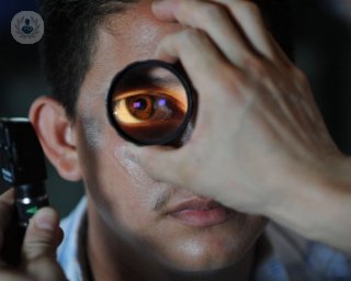
- Ophthalmoscopy: Examines that back of the eye, including the retina.
- Refraction test: Used to verify prescription glasses.
- Verification of chromatic vision.
- Visual acuity test: Determines the smallest letters that can be read on a standardised chart (Snellen chart).
- Slit lamp test: Checks the structures in the front of the eye.
- Eye ultrasound.
What causes it?
The main causes for retinal detachments are due to retinal tears or holes.
Other causes include:
- Ageing can cause retinal thinning and deterioration.
- Vitreous retraction: The vitreous is attached to the retina by many fibres. If it retracts, it may cause a tear or a hole. This is the most common cause of retinal detachment.
- Abnormal eye growth: Vitreous contraction may be caused due to myopia, inflammation or swelling, or eye trauma.
How can it be prevented?
It is crucial to have an ophthalmologic examination with pupillary dilation to be to check the retinal periphery.
If degenerative lesions that could cause detachment are found, laser treatment will be recommended.
Day-to-day tips:
- Wear protective glasses to prevent eye trauma.
- Diabetic patients should carefully monitor their blood sugar.
- See an ophthalmologist once a year.
- Be aware of new flashing light or floater symptoms.
Early diagnosis and treatment are essential for a good outcome.
What is the treatment?
In most cases treatment will involve surgery, for example in both rhegmatogenous and tractional retinal detachment. The former is the most common type of retinal detachment.
In cases of serious retinal detachment, the cause needs to be treated. This may be done by surgery, medications, or radiotherapy, amongst other treatments.
There are different types of surgery that can be used, depending on each case. Currently, the most commonly used is microsurgery through posterior vitrectomy via pars plana.
The aim of the treatment is to:
- Close the tear or rupture what has caused retinal detachment using laser or cryotherapy.
- Put the retina back in its place.
The procedure is normally done on an outpatient basis. Local anaesthetic and sedation are used.


Is seeing flashing lights in your eye a sign of retinal detachment?
By Miss Miriam Minihan
2024-07-27
Flashing lights in your eyesight look like flashes of lightening or if someone had taken a photograph with using the flash. Are you seeing them frequently and wondering if they are a cause for concern? Here, expert ophthalmologist Miss Miriam Minihan explains whether they are a sign of an underlying problem or not. See more

What is a detached retina and how does it happen?
By Mr Mahi Muqit
2024-07-26
Consultant ophthalmologist Mr Mahi Muqit discusses how to treat a detached retina, which occurs when the thin lining at the back of the eye starts to pull away from the blood vessels that supply it with oxygen and nutrients. See more

How does intravitreal injection therapy work?
By Miss Sudeshna Patra
2024-07-26
Here, extremely experienced and qualified consultant ophthalmic surgeon, Miss Sudeshna Patra is on hand here in this article to provide all the key nuggets of information regarding intravitreal injection therapy for eye retinal conditions. See more


5 eye conditions you should know about
By Mr Praveen Patel
2024-07-26
Top ophthalmologist Praveen Patel gives us a run-down of 5 eye conditions you should know about, from macular degeneration to diabetic retinopathy! See more
Experts in Retinal detachment
-

Mr Mahi Muqit
OphthalmologyExpert in:
- Eye floaters
- Epiretinal membrane
- Diabetic retinopathy
- Vitrectomy
- Macular hole
- Retinal detachment
-

Professor James Bainbridge
OphthalmologyExpert in:
- Cataracts
- Epiretinal membrane
- Macular hole
- Retinal detachment
- Eye floaters
- Retinal detachment surgery
-

Professor Tom Williamson
OphthalmologyExpert in:
- Retinal detachment
- Macular hole
- Epiretinal membrane
- Diabetic retinopathy
- Cataracts
- Vitrectomy
-

Mr Hadi Zambarakji
OphthalmologyExpert in:
- Retinal detachment
- Macular hole
- Epiretinal membrane
- Macular degeneration (AMD)
- Diabetic retinopathy
- Cataracts
-

Mr Roger Wong
OphthalmologyExpert in:
- Retina
- Diabetic retinopathy
- Retinal detachment
- Macular hole
- Epiretinal membrane
- Macular degeneration (AMD)
- See all

The Meriden Hospital - part of Circle Health Group
The Meriden Hospital - part of Circle Health Group
Clifford Bridge Road, Coventry
No existe teléfono en el centro.
By using the telephone number provided by TOP DOCTORS, you automatically agree to let us use your phone number for statistical and commercial purposes. For further information, read our Privacy Policy
Top Doctors
Guy’s and St Thomas’ Private Healthcare
Guy’s and St Thomas’ Private Healthcare
Guy’s Hospital, Great Maze Pond
No existe teléfono en el centro.
By using the telephone number provided by TOP DOCTORS, you automatically agree to let us use your phone number for statistical and commercial purposes. For further information, read our Privacy Policy
Top Doctors
The Alexandra Hospital - part of Circle Health Group
The Alexandra Hospital - part of Circle Health Group
Mill Ln, Cheadle
No existe teléfono en el centro.
By using the telephone number provided by TOP DOCTORS, you automatically agree to let us use your phone number for statistical and commercial purposes. For further information, read our Privacy Policy
Top Doctors
-

The Meriden Hospital - part of Circle Health Group
Clifford Bridge Road, Coventry , CoventryExpert in:
- Cancer
- Cardiology
- Colorectal surgery
- Shoulder surgery
- Hand surgery
- General Surgery
-

Guy’s and St Thomas’ Private Healthcare
Guy’s Hospital, Great Maze Pond, SE1 South Bank LondonExpert in:
- Allergy
- Cardiology
- General Surgery
- Maxillofacial Surgery
- Thoracic Surgery
- Maternity care
-

The Alexandra Hospital - part of Circle Health Group
Mill Ln, Cheadle, CheadleExpert in:
- Hip
- Cardiology
- Shoulder and elbow
- Paediatrics
- Foot and ankle
- Knee
- See all
- Most viewed diseases, medical tests, and treatments
- Orthoptics
- Medicolegal
- Dermal fillers
- Headache
- Strabismus (squint)
- Glaucoma
- Diplopia (double vision)
- Amblyopia
- Vision therapy
- Laser photocoagulation