A comprehensive guide to MRIs
Written by:MRIs are extremely common, used to analyse parts of the body and often to diagnose tumours. Esteemed consultant cardiologist with cardiac MRI expertise Dr Teresa Castiello provides a guide, answering all your commonly asked questions.
What is an MRI?
An MRI, or magnetic resonance imaging, is a medical imaging technique that uses a powerful magnetic field and radio waves to create detailed images of the body's internal structures. It provides a non-invasive way to visualise organs, tissues, and structures in great detail, in-depth helping doctors diagnose and monitor various medical conditions.
What is a GBCA in MRI? Is it safe?
GBCA (gadolinium-based contrast agent) is a type of dye used in some MRI scans in order to enhance the visibility of certain structures or abnormalities. GBCAs are generally safe and well-tolerated. However, in rare cases, they can cause a condition called nephrogenic systemic fibrosis (NSF) in individuals with severe kidney problems. If you have any concerns about the safety of GBCAs, it is important to discuss them with your doctor before undergoing an MRI.
When might a doctor suggest an MRI?
A doctor may suggest an MRI when they need more detailed information about a particular area of the body that cannot be obtained through other imaging techniques. MRI is commonly used to evaluate conditions affecting the brain and spinal cord, joints, soft tissues, and organs such as the liver, kidneys, and heart.
MRI is particularly useful in diagnosing conditions such as tumours, injuries, infections, and abnormalities of the musculoskeletal and nervous systems. Neuroradiologists such as Dr Francesco Carletti and Dr Felice D’Arco have extensive training to report neurology studies respectively in adults and children.
A cardiac MRI gives accurate information about the heart structure, function and scar pattern that the heart muscle may have for a wide number of underlying conditions. MRI cardiologists such as Dr Giulia Benedetti are specifically trained to carry out and report those scans.
How can I prepare for an MRI?
Preparing for an MRI typically involves a few simple steps. You may be asked to remove any metallic objects, such as jewellery or clothing with zippers, as the MRI uses a strong magnetic field. Before having an MRI, you should inform your doctor if you have any metal implants, devices, or pacemakers, as they may affect your ability to undergo an MRI. In some cases, you may need to fast for a few hours before the scan, especially if you are having an abdominal or pelvic MRI.
Your doctor will provide you with specific instructions based on your individual case. If you need to have an adenosine-stress cardiac MRI you need to refrain from caffeine and theanine for at least 12 hours.
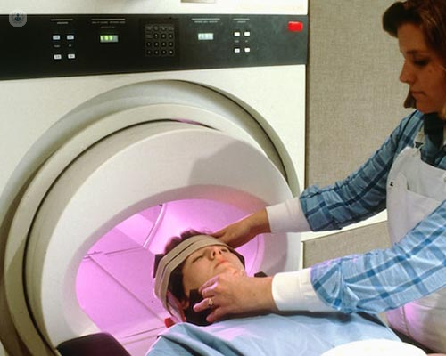
What happens during an MRI?
During an MRI, you will lie down on a movable table that slides into the MRI machine. It is important to remain as still as possible during the procedure to obtain clear images. The machine will generate a series of loud knocking or buzzing sounds, but you will be provided with earplugs or headphones to minimise the noise. The technologist will be able to communicate with you through an intercom system. The scan can take anywhere from 15 minutes to over an hour, depending on the area being scanned. In some cases, a contrast agent may be injected into a vein to enhance the visibility of certain structures.
What is the difference between MRI and CT scan?
While both MRI and CT scans are imaging techniques, they differ in how they generate images and the type of information they provide. CT, or computed tomography, uses X-rays to create cross-sectional images of the body. It is particularly useful for imaging bones, detecting fractures, and evaluating acute conditions like bleeding or trauma.
MRI, on the other hand, uses a magnetic field and radio waves to produce detailed images of soft tissues, organs, and the nervous system. It is especially valuable for visualising the brain, spinal cord, joints, and soft tissues. MRI does not use ionizing radiation such as CT scans, making it safer for repeated imaging and for certain populations, such as young people, or children or those patients who require regular interval scans.
If you would like to book a consultation with Dr Castiello, do not hesitate to do so by visiting her Top Doctors profile today.