Echocardiogram – Part 2: The procedure
Written by:Our hearts are a vital part of our bodies, essential to keeping us alive. When heart problems occur, or even just during health screenings, your doctor may perform an echocardiogram to examine your heart health. In the second part of his series on echocardiograms, top cardiologist Dr Allan Harkness explains the echocardiogram procedure.

What will I be asked to do during the echocardiogram?
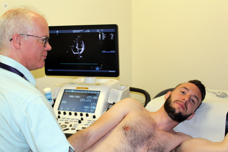
To look at the heart from all angles, the sonographer will need to place the ultrasound probe directly on your skin at several places. These are:
- Over the front of your chest
- Under your left breast
- At the base of your neck
- Just under your ribs at the front
To get good pictures, the probe needs to be covered in a special lubricant gel.
Therefore, to have an echocardiogram, you need to take your clothes off from the waist up – including any bra. You will be offered a gown to wear instead. This gown is worn like a cape, opening at the front.
During the scan you will be asked to lie on a special couch which has the head tilted up. For most of the time, you will be lying on your left hip but for some pictures, you will be asked to lie on your back or right hip. The sonographer may ask you to breathe in or out or hold your breath at times. This helps move the lungs out of the way to get the clearest pictures.
After the scan, you will be given paper towels to wipe off all the gel. If you miss some when you put your clothes back on, it may leave a sticky mark, but this should wash out.
An echocardiogram takes from 30 to 60 minutes to do. You can carry on with your day as normal afterwards.
How can I prepare for an echocardiogram?
You should try to wear clothes that are easy to take off with minimum layers and buttons. Avoid a top that is dry-clean only and leave any necklace or chains at home. You will be lying for a while on your left hip and sometimes your right hip, so put coins, car keys and phones in a jacket or purse.
For a normal echocardiogram, you do not need to do anything different from your normal routine. You should take all your usual medicines and you don’t need to miss any meals.
If you need help to get about or help with dressing, you might want a family member or friend to come with you. They can usually come in with you during the study or else sit outside – it is up to you. If you want a chaperone with you, just let the receptionist know beforehand.
There are some special forms of echo where it is important to fast or avoid some medication beforehand so always check your appointment card for special instructions or ask if you are not sure.
What happens after the results are received from an echocardiogram?
It can take a while to fully analyse an echo study after all the pictures are taken. A final report will be written and sent to your cardiologist.
Your cardiologist will review the report and images taken and put these into context with the clinical question being asked.
Usually the results will be given to you at your next follow up appointment. If the scan was expected to be normal and this turned out to be the case, you might prefer to agree on the results being sent to you by letter. However, you can always make an appointment if you need to ask more questions.
Sometimes, deciding the right thing to do for a heart condition can be very complicated. This is more often the case with heart valve disease. In these situations, a multidisciplinary team of experts may need to meet to discuss your case. This team would include your cardiologist and other cardiologists with different areas of expertise together with cardiac surgeons.
Your cardiologist would then meet with you to present you with the options discussed at the team meeting and help you decide what treatment you wish to have.
In part 3, Dr Allan Harkness describes more advanced forms of echo and what additional imaging tests may be required.


