4D ultrasound

Professor Nazar Amso - Obstetrics & gynaecology
Created on: 04-29-2014
Updated on: 05-20-2015
Edited by: Conor Lynch
What is 4D ultrasound?
Ultrasound is an imaging technique which allows a foetus to be observed in real time in the womb. As well as providing a high-quality image of the foetus, 4D ultrasound can be used for diagnostic imaging, allowing a healthcare specialist to observe the body's structures and tissues.
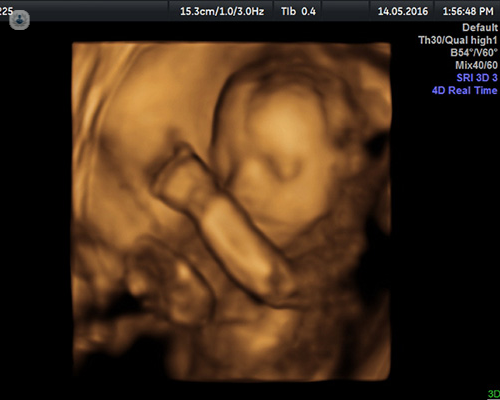
During pregnancy, 3D ultrasound allows you to see three-dimensional images of your baby in the womb (as opposed to the traditional 2D images), while with 4D technology the image is constantly updated in real time and you can see it moving in the form of a video.
How is a 4D ultrasound scan performed?
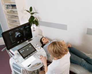
4D ultrasound, like other ultrasound scans, uses high-frequency sound waves to generate images of organs and tissues inside the body. These sound waves are emitted by a probe or transducer, which is passed over the region to be scanned by the doctor.
During pregnancy, this test is performed to observe the status and development of the foetus without exposing it to radiation. The doctor administers a small amount of cold gel on the belly of the pregnant woman, and uses a probe to capture the images of the uterus and project them on a screen.
How do 4D ultrasound scans differ from more traditional scanning?
Traditional ultrasound scans produce a flat 2D image, but modern technology can combine data from scans taken by the probe from different angles to create a 3D image. When this 3D image is refreshed in real time, it results in a live video known as a 4D ultrasound.
What is the aim of 4D ultrasound scanning?
Ultrasound scans are very important for many parents, since they not only serve to monitor the development of the baby, but also to show the first images of the foetus, which can be retained as a memory or souvenir.
The advantage of 4D ultrasound is that in showing the developing baby in three dimensions and in motion, it is much easier to see the baby clearly. This makes it easier for doctors to diagnose any developmental problems, such as a cleft lip, which may be harder to spot on a standard 2D ultrasound.
What can be seen on a 4D ultrasound scan?
A 4D ultrasound scan can give information about a baby’s development, including size, heart rate and gender, as well as allowing parents to see their baby in detail and give reassurance that all is well.
During the scan, it is possible to see the baby in real time and perhaps smile, yawn, or open and close their eyes, for example. Importantly, however, 4D ultrasound scans are primarily a medical test, where a qualified specialist can advise on your baby’s health and development.
They also allow the healthcare practitioner to observe if the foetus’ organs are developing well and if any birth defects, such as cleft palate, are present. The estimated weight can also be established as well as the levels of amniotic fluid, giving a vital assessment about the health of the pregnancy.
When is the best time to undergo one?
The best time for a 4D ultrasound scan is usually between weeks twenty four and thirty of pregnancy. At this stage, the baby is fairly well-developed, but has not yet grown so large that it cannot be seen well on the scan.
How does it feel during a 4D ultrasound scan?
Ultrasound is a totally painless non-invasive imaging technique, both for the mother and baby. The scan is completely safe as it uses sound waves to look inside the body, rather than penetrating forms of radiation, such as those used in X-rays.
Which specialist performs a 4D ultrasound scan?
Specialists in obstetrics perform 4D ultrasound scans. It is important to seek out a registered and qualified specialist to perform a 4D ultrasound scan as high-street scans aimed at giving parents a “keep-sake” may not provide reliable information about your baby’s development and health.

What are the different types of ultrasound scans in pregnancy?
By Dr Srividhya Sankaran
2024-07-27
Different types of ultrasound are undertaken depending on the stage of the pregnancy, and also for various reasons. These can include calculating the chances of the pregnancy being affected with Down’s syndrome and risk of going into preterm labour, alongside many others. Highly-experienced consultant in maternal-foetal medicine (MFM) and obstetrics Dr Srividhya Sankaran, is here to explain these different ultrasound scans. See more
Experts in 4D ultrasound
-

Miss Luxmi Velauthar
Obstetrics & gynaecologyExpert in:
- Fetal medicine
- Premature birth
- Recurrent miscarriage
- Menstrual disorders
- Pre-eclampsia
- 4D ultrasound
-
Professor Nazar Amso
Obstetrics & gynaecologyExpert in:
- 4D ultrasound
- Assisted reproductive technology
- Hysteroscopy
- Menstrual disorders
- Pelvic pain
- Transvaginal ultrasound
- See all

Oak Tree Clinic
Oak Tree Clinic
2A, Oak Tree Court Mulberry Drive, Cardiff Gate Business Park, Cardiff
No existe teléfono en el centro.
By using the telephone number provided by TOP DOCTORS, you automatically agree to let us use your phone number for statistical and commercial purposes. For further information, read our Privacy Policy
Top Doctors
LycaHealth Canary Wharf
LycaHealth Canary Wharf
1 Westferry Circus, Canary Wharf. E14 4HD
No existe teléfono en el centro.
By using the telephone number provided by TOP DOCTORS, you automatically agree to let us use your phone number for statistical and commercial purposes. For further information, read our Privacy Policy
Top Doctors
-

Oak Tree Clinic
2A, Oak Tree Court Mulberry Drive, Cardiff Gate Business Park, Cardiff, CardiffExpert in:
- Ultrasound
- In vitro fertilisation (IVF)
- Fertility
- Obstetrics and Gynaecology
- Aesthetic Medicine
- Health check up
-

LycaHealth Canary Wharf
1 Westferry Circus, Canary Wharf. E14 4HD, Central LondonExpert in:
- Cardiology
- Dermatology
- Diagnostic Imaging
- Women’s health
- Most viewed diseases, medical tests, and treatments
- Platelet-rich plasma
- Medicolegal
- Early menopause
- Perimenopause
- Premature menopause
- Premature ovarian insufficiency (POI)
- Peritoneal carcinomatosis
- Pre-eclampsia
- Women's health
- Robotic surgery