Chest X-ray
Dr Sundeep Kaul - Pulmonology & respiratory medicine
Created on: 04-13-2018
Updated on: 03-21-2023
Edited by: Sophie Kennedy
What is a chest X-ray?
Chest X-rays may be performed for a number of reasons. An x-ray is a quick, painless and effective imaging test that uses low bursts of ionising radiation to produce pictures of the inside of the body. When performed on the chest, it allows the doctor to examine the heart, lungs, bones, airways, and blood vessels in the chest.

What does it involve?
X-rays are a form of radiation like light or radio waves, and are able to pass through most objects, including the body. However, different parts of the body absorb different amounts of X-rays, for example, bone absorbs much more than soft tissue.
An X-ray machine is used to emit these X-rays in small bursts at the patient’s chest. The X-rays pass through the body, and create an image when they hit a special image recording plate placed on the other side of the patient.
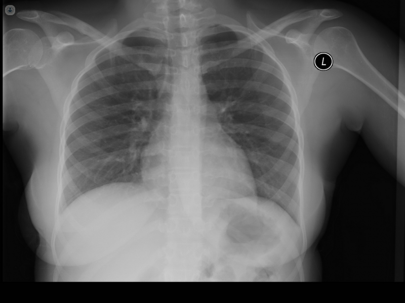
The image will depict the inside of the chest, displaying different tissues and organs in different shades. For example, bones, such as the ribs, will appear white as they block and absorb most of the x-rays, preventing them from reaching the film/plate, while soft tissue, such as the lungs, appear in different shades of grey, and air appears black.
What is a chest X-ray for?
A chest X-ray may be needed to assess the extent of the damage to the chest following an accident or injury, to monitor the progression of diseases, like cystic fibrosis, or to examine the heart.
A doctor may also order a chest X-ray if his patient is suffering the following symptoms:
- Chest pain
- Shortness of breath
- Persistent cough
- Fever
These symptoms can be related to conditions involving the organs in the chest, and the chest X-ray can help the doctor to diagnose a number of these conditions, including:
- Broken or fractured ribs
- Emphysema
- Heart failure
- Lung cancer
- Pneumonia
- Pneumothorax (a build-up of air between the lungs and the chest wall)
It can also be useful in the positioning of medical devices in the chest.
How can you prepare for it?
There is no special preparation required for a chest X-ray. The patient may need to remove jewellery, glasses, certain dental appliances, and certain items of clothing (particularly those with metal elements).
X-rays are generally not performed on pregnant women, so as not to expose the foetus to radiation, so it is important to inform your doctor if there is a chance you are pregnant.
What does it feel like during the procedure?
The patient stands against the image recording plate, while a technologist puts them in the correct position. Usually, images are taken from both the back and the side. For the former, the patient must stand with hands on hips and their chest pressed against the plate, while in the latter case, the patient raises their hands while pressing their side against the plate. While the images are taken, the patient must remain still, and may have to hold their breath for a few seconds to keep their chest from moving.
A chest X-ray is a quick and painless procedure, although the patient may experience some discomfort from the cold plate.
Chest X-ray results
The results may be ready for review immediately, although the doctor will often examine them before discussing them with the patients.
While the X-ray itself can sometimes be sufficient for a diagnosis, in other cases, a follow-up exam may be needed. This can be for a number of reasons. In the case of an abnormal chest X-ray, other special imaging techniques may be required.


