Densitometry

Dr Mymoona Alzouebi - Clinical oncology
Created on: 06-20-2013
Updated on: 04-26-2023
Edited by: Sophie Kennedy
What is densitometry?
Bone densitometry refers to special scanning techniques used to visualise bone structure and analyse bone mineral density (BMD). As opposed to other X-ray techniques, densitometry emphasises quantification of the bone, with a shorter scanning time and precise results, allowing doctors to monitor bone loss in patients. The standard method of bone densitometry is the dual-energy x-ray absorptiometry (DEXA/DXA) scan.
What does bone densitometry involve?
Densitometry involves firing small bursts of ionising radiation through the patient to create an image of inside their body. The bones absorb part of this radiation, and the amount absorbed reflect the density of the bones, which will be observable on the image.
Early densitometry techniques, such as single-photon absorptiometry and later dual-photon absorptiometry used iodine-125 and gadolinium-153 respectively, while modern DXA scans use low-level X-rays.
Why is densitometry done?
The main use of DXA is to diagnose and monitor the progress and/or treatment of osteoporosis. Bone is constantly being broken down and built up again by the body, but as we get older, more is broken down than is formed, and various conditions or circumstances can increase the rate of this breakdown. Osteoporosis is the result of this – a condition where the hollow spaces in the bones become wider and the bones become brittle as a result.
Preparation for a DXA scan
As with most X-rays, little to no preparation is required. You should remove jewellery before the DXA scan and it is advisable to wear loose, comfortable clothes. You should not take any calcium supplements in the twenty-four hours before the test and inform your doctor and technologist if have recently had a test involving contrast material (e.g. for a CT scan), a radioisotope, or a barium scan. You should also let them know if there is any chance you could be pregnant.
What to expect during a DXA scan
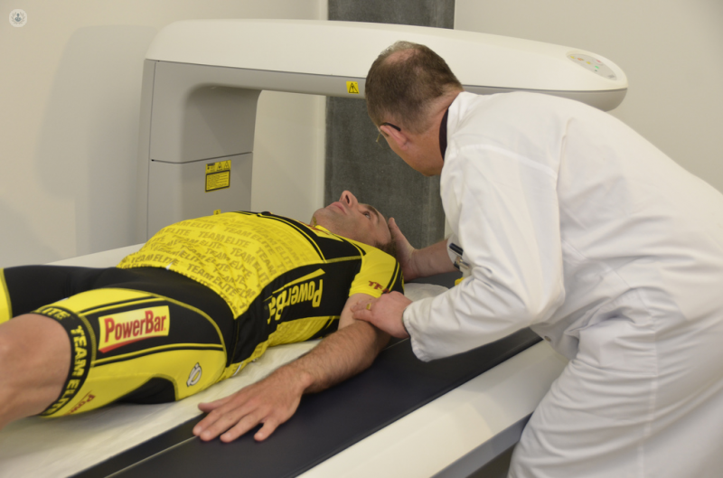
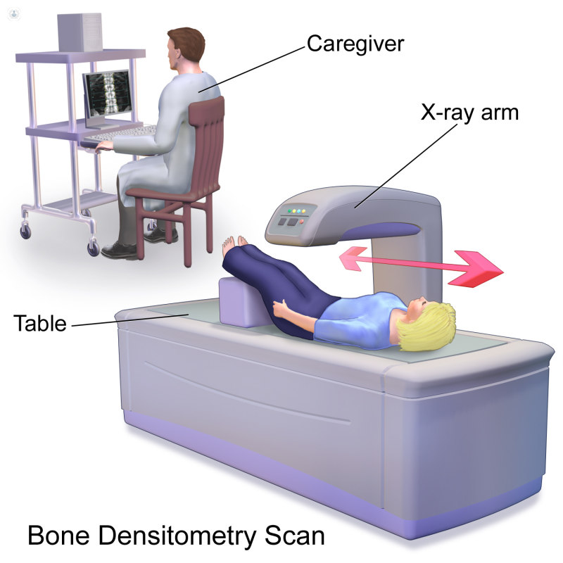
Densitometry scans are quick and painless. The patient usually lies on a table with the X-ray generator beneath them and the image generator above them. They will be asked to lie still and hold their breath for a few seconds if the torso is being scanned to avoid blurring the images. Patients may need follow-up scans months or years later to monitor their progress.
- Most viewed diseases, medical tests, and treatments
- DEXA scan
- Back pain
- Medicolegal
- Peritoneal carcinomatosis
- Family history of breast cancer
- Skin biopsy
- Cephalometric
- Stereotactic radiotherapy
- Hyperthermia therapy
- Endovascular brachytherapy