Dermoscopy
Dr Cristina Bordea - Dermatology
Created on: 06-29-2015
Updated on: 04-03-2023
Edited by: Jay Staniland
What is dermoscopy?
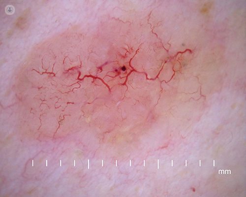
Dermoscopy, which is also known as dermatoscopy, is a skin examination technique that uses a dermatoscope. A dermatoscope has a high quality magnifying lens which allows careful examination of the skin structure, and can be used to diagnose skin cancer and to map moles. A dermatoscope is a hand-held device that takes high-resolution photos.
What does dermoscopy involve?
Using a dermoscope, skin lesions are evaluated in terms of their pigmentation, colours and structure (symmetry, uniformity, border shape). Evaluating these characteristics can aid in the diagnosis of the following:
- Melanoma
- Benign moles
- Pigmented basal cell carcinoma
- Haemangioma
Dermoscopy may also be used to examine the skin for the following:
Why is dermoscopy done?
When used by an expert, dermoscopy can be effective in identifying melanomas and confirming malignant skin cancers. This can reduce the number of unneeded excisions of benign lesions. Identifying melanomas by the naked-eye is not accurate, and using a dermoscope improves this. Dermoscopy can also be used for patients who have many melanocytic nevi (moles) that do not meet the criteria of a melanoma, but look suspicious. Hence, a dermoscopy can track any changes in these and determine whether or not a biopsy is required.
What to expect during a dermoscopy:
During a dermoscopy, a special liquid gel is applied to the area of interest and the dermoscope is placed onto the lesion for examination. This gel allows the clinician to see structures below the surface of the skin. Newer models of dermoscope have eliminated the need for using the gel. There is no discomfort felt during a dermoscopy.
What do abnormal results mean?
If malignancies are detected, treatment can be planned which will involve biopsy and excision.


Keeping an eye on your moles: Detecting melanoma early on
By Dr Ben Esdaile
2024-07-26
In the UK, there are around 15,000 new cases of melanoma every year, making it the fifth most common cancer in the UK. Dr Ben Esdaile is a leading dermatologist and an expert in the early diagnosis and treatment of skin cancer. Find out from an expert how you can both prevent melanoma, as well as detect it in its early stages. See more
Experts in Dermoscopy
-

Dr Kapil Bhargava
DermatologyExpert in:
- Skin cancer
- Alopecia
- Moles
- Dermoscopy
- Mohs surgery
- Cosmetic dermatology
-

Dr Ben Esdaile
DermatologyExpert in:
- Psoriasis
- Acne
- Skin cancer
- Paediatric dermatology
- Nail diseases
- Dermoscopy
-
Dr Cristina Rodriguez-Garcia
DermatologyExpert in:
- Dermoscopy
- Psoriasis
- Skin cancer diagnosis
- Paediatric dermatology
- Acne
- Eczema
-
Dr Cristina Bordea
DermatologyExpert in:
- Skin cancer
- Scar revision
- Moles
- Melanoma
- Dermoscopy
- Clinical dermatology
-
Dr Rubeta Matin
DermatologyExpert in:
- Cryotherapy
- Dermoscopy
- Lymphoma
- Melanoma
- Photodynamic therapy
- Skin cancer diagnosis
- See all

Derma Reading
Derma Reading
Shepherds Hill, Woodley, RG6 1FE
No existe teléfono en el centro.
By using the telephone number provided by TOP DOCTORS, you automatically agree to let us use your phone number for statistical and commercial purposes. For further information, read our Privacy Policy
Top Doctors
The Chiltern Hospital - part of Circle Health Group
The Chiltern Hospital - part of Circle Health Group
London Rd, Great Missenden HP16 0EN
No existe teléfono en el centro.
By using the telephone number provided by TOP DOCTORS, you automatically agree to let us use your phone number for statistical and commercial purposes. For further information, read our Privacy Policy
Top Doctors
Skin Inspection Dermatology Clinic
Skin Inspection Dermatology Clinic
Harley Street Clinic 55 Harley Street 4th Floor, London
No existe teléfono en el centro.
By using the telephone number provided by TOP DOCTORS, you automatically agree to let us use your phone number for statistical and commercial purposes. For further information, read our Privacy Policy
Top Doctors
-
Derma Reading
Shepherds Hill, Woodley, RG6 1FE, ReadingExpert in:
- Acne
- Skin Cancer
- Paediatric Dermatology
- Moles
- Aesthetic Medicine
- Hair loss
-
The Chiltern Hospital - part of Circle Health Group
London Rd, Great Missenden HP16 0EN, Great MissendenExpert in:
- Allergies Ophthalmological
- Clinical analysis
- Cancer
- Breast Cancer
- Skin Cancer
- Prostate Cancer
-
Skin Inspection Dermatology Clinic
Harley Street Clinic 55 Harley Street 4th Floor, London, W1G Marylebone LondonExpert in:
- Skin Cancer
- Clinical Dermatology
- Surgical Dermatology
- Diagnosis of Cancer
- Moles
- Cancer Treatment
- See all
- Most viewed diseases, medical tests, and treatments
- FUE hair transplant
- Weight loss injections
- Carpal tunnel syndrome
- Platelet-rich plasma
- Anti-ageing treatments
- Medicolegal
- Skin biopsy
- Cleft palate
- Herpes
- Genital dermatology