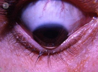
Keratoconus

Mr Radwan Almousa - Ophthalmology
Created on: 11-13-2012
Updated on: 05-20-2015
Edited by: Jay Staniland
What is keratoconus?
Keratoconus is an eye disorder in which the corneal tissue thins and deforms. The cornea is unable to support the internal eye pressure and it bulges out in a cone shape, hence the name, kerato, which means cornea, and conus, which means cone shaped in Latin.
It is a degenerative condition of the cornea that appears progressively in one or both eyes.
Disease prognosis
Although keratoconus is a degenerative disease, it can be corrected with glasses, with contact lenses, and different surgical procedures.
A patient with keratoconus should have regular corneal topographies to detect changes in the cornea.
What are the symptoms of keratoconus?
Keratoconus usually affects young people in the form of myopia and astigmatism as well as blurred vision. It may also cause eye irritation and increased photophobia (light sensitivity). A specialist will confirm the disease when and if they detect corneal thinning in the central or paracentral part of the cornea.
Tests for keratoconus
A corneal topography is done to check the curvature of the front and the back of the cornea and to detect areas of corneal thinning. New methods also measure the thickness of the very superficial layer of the cornea that is called the epithelium, as it starts to have different behaviour in keratoconus patients.
What causes it?
Keratoconus was thought to be a pure degenerative disease, however, new evidence indicates the disease to be part of an inflammatory condition, with intracorneal inflammatory cells causing focal thinning and protrusion of the cornea. This is evident as the disease is more common in patients with a history of allergic conditions such as atopic dermatitis and asthma. Bad habits associated with allergic conditions, like eye rubbing, are known risk factors. Genetics may also play a role, as the disease is more common in some genetic conditions such as Down's syndrome.
How can it be prevented?
To prevent keratoconus, it is recommended that you have your eyes checked at least once a year. There are also some actions that are best avoided, such as continuously rubbing your eyes. Early detection is crucial to be able to treat the symptoms and prevent corneal degeneration.
What is the treatment?
Prevention is the first thing to consider, especially if the patient feels that their glasses prescription is changing in a short period of time, as mild cases could be managed with glasses and contact lenses.
If the ophthalmologist discovers the keratoconus in an early stage, it could be controlled with collagen cross linking, which involved stiffening the cornea with ultraviolet light and vitamin B (Riboflavin).
In more advanced cases in which the patient cannot tolerate contact lenses, and if the vision is not corrected enough with glasses or contact lenses, a new procedure involving implanting rings inside the cornea (CAIRS surgery). The rings are made from a donor cornea, which makes them safer than the plastic rings that were previously used to correct the keratoconus corneas.
A new procedure to add tissue to severe cases of keratoconus to increase the corneal thickness and to flatten the cornea has been introduced in a few centres around the world.
Eventually a partial thickness corneal graft can be considered in severe cases to restore the clarity of the cornea.
What specialist should I see?
Keratoconus should be managed by an ophthalmologist.

How the latest keratoconus treatment can halt progression and correct vision
By Mr Kieren Darcy
2024-10-31
Keratoconus has a progressive nature, and patients who have this ophthalmological condition can experience issues with quality of focus and vision. Sight can be severely affected when it’s in its late stages, too. Here to explain why it’s important to detect it as soon as possible as well as explain the latest treatment, is Mr Kieren Darcy, a leading consultant eye surgeon and ophthalmologist in Bristol. See more


How serious is keratoconus?
By Mr CT Pillai
2024-10-31
Keratoconus is a condition that affects the front part of the eye, causing it to bulge outwards in an irregular cone shape and results in blurry vision. Being diagnosed with an eye condition that you may not have heard of before can be pretty scary. We've asked one of our top ophthalmologists Dr CT Pillai to explain just how serious it is. See more


I have keratoconus, will I lose my sight?
By Mr Arun Brahma
2024-10-31
Keratoconus primarily affects the cornea and is a condition that means the cornea has become cone-shaped. Naturally, as with any eye condition, patients worry that they'll lose their sight. Mr Arun Brahma explains what you should know about keratoconus and preserving your sight. See more

Keratoconus: a comprehensive guide
By Mr Don Williams
2024-10-28
Keratoconus is an eye condition where the cornea progressively thins and bulges into a cone shape. It distorts vision, often requiring special contact lenses or surgery for correction. Here to explain more about the condition is reputable consultant specialist optometrist Mr Don Williams, who provides us with a comprehensive guide. See more
Experts in Keratoconus
-

Miss Valerie Saw
OphthalmologyExpert in:
- Cataracts
- Laser eye surgery
- Dry eye
- Keratoconus
- Corneal transplant
- Corneal cross-linking
-

Mr Amit Patel
OphthalmologyExpert in:
- Cataracts
- Refractive lens exchange
- Laser eye surgery
- Corneal transplant
- Keratoconus
- Dry eye
-

Mr Connan Tam
OptometryExpert in:
- Orthoptics
- Keratoconus
- Gas permeable contact lenses
- Dry eye
- Myopia
- Visual stress (Meares-Irlen syndrome)
-

Mr Mohamed Elalfy
OphthalmologyExpert in:
- Cataracts
- Laser eye surgery
- Dry eye
- Eye examination
- Keratoconus
- YAG laser capsulotomy
-

Mr Aris Konstantopoulos
OphthalmologyExpert in:
- Cataracts
- Laser eye surgery
- Keratoconus
- Corneal transplant
- Corneal cross-linking
- Lens replacement (intraocular lenses)
- See all

Laser Vision
Laser Vision
Prema Compass Road North Harbour Business Park Portsmouth PO6 4RP
No existe teléfono en el centro.
By using the telephone number provided by TOP DOCTORS, you automatically agree to let us use your phone number for statistical and commercial purposes. For further information, read our Privacy Policy
Top Doctors
Tunbridge Wells Eye Centre
Tunbridge Wells Eye Centre
7 Vale Ave, Tunbridge Wells TN1 1DJ
No existe teléfono en el centro.
By using the telephone number provided by TOP DOCTORS, you automatically agree to let us use your phone number for statistical and commercial purposes. For further information, read our Privacy Policy
Top Doctors
Dry Eye Centre
Dry Eye Centre
7 Devonshire St, London W1W 5DY, United Kingdom
No existe teléfono en el centro.
By using the telephone number provided by TOP DOCTORS, you automatically agree to let us use your phone number for statistical and commercial purposes. For further information, read our Privacy Policy
Top Doctors
-
Laser Vision
Prema Compass Road North Harbour Business Park Portsmouth PO6 4RP, HavantExpert in:
- Cataracts
- Laser eye surgery
- ICL lens implants
- Ophthalmology
- Keratoconus
- Lens replacement
-

Tunbridge Wells Eye Centre
7 Vale Ave, Tunbridge Wells TN1 1DJ, Tunbridge WellsExpert in:
- Cataracts
- Oculoplastics surgery
- Laser eye surgery
- Refractive surgery
- Facial aesthetics
- Ophthalmology
-

Dry Eye Centre
7 Devonshire St, London W1W 5DY, United Kingdom, West LondonExpert in:
- Contact lenses for dry eyes
- Dry eye
- Optometry
- See all
- Most viewed diseases, medical tests, and treatments
- Minimal access surgery (keyhole surgery)
- Botulinum toxin (Botox™)
- Orthoptics
- Medicolegal
- Dermal fillers
- Headache
- Strabismus (squint)
- Glaucoma
- Diplopia (double vision)
- Amblyopia