Prostate MRI

Mr Christof Kastner - Urology
Created on: 04-17-2018
Updated on: 05-09-2023
Edited by: Aoife Maguire
What is a prostate MRI?
An MRI scan creates images using radio waves an magnetic fields. An MRI of the prostate gland can help identify any abnormalities, including prostate cancer, infections or benign prostatic hyperplasia (BPH). Unlike X-rays, MRI scans do not use ionising radiation. The images produced from a prostate MRI provide a high level of detail to aid in a diagnosis or evaluation.
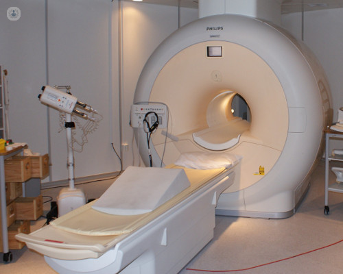
What does a prostate MRI involve?
The MRI machine is a large tube, with a bed that slides in and out of it. During the scan you lie on the bed on your back, and straps may be used to help you keep still. An endorectal coil, which is a thin wire covered with a latex balloon which is placed inside your rectum might be used during the MRI. If so, it is inserted using lubrication and it allows a more detailed image of the prostate to be generated. You might also be given a contrast material which is inserted intravenously through a catheter in your hand or arm. Contrast material can help show the body’s anatomy better. Once you are inside the MRI machine, scans commence which take a few minutes at a time. A number of scans will be taken, and the whole process is usually completed within 45 minutes.
What is a prostate MRI for?
The most common use of a prostate MRI is to check for prostate cancer, or to determine the stage of prostate cancer. A prostate MRI can also be carried out to check for:
- infection or an abscess
- enlarged prostate (BPH)
- surgical complications
- congenital abnormalities
How can you prepare for a prostate MRI?
Before a prostate MRI it is important to confirm whether or not you have any of the following (this is because MRIs use magnetism):
- surgical pins
- pacemaker
- cochlear implants
- any metal fragments in your body
It may also be necessary to stop eating and drinking an hour or so before having the MRI scan. It is also important to remove any jewellery, watches, hair clips or coins from yourself or your clothes before the scan.
What does it feel like during a prostate MRI?
Although painless, sometimes it is challenging to remain still for the duration of the MRI scan. It can also feel warm during an MRI scan. Some people find an MRI a bit claustrophobic too. At certain points in a scan, it might be necessary to hold your breath for a few seconds.
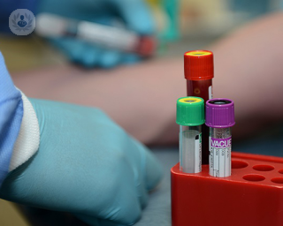
What would a “bad” result mean?
Once the scans are complete, a radiologist will interpret the results and create a report for your specialist. Results are usually available in one to two weeks following the scan. A “bad” result might indicate prostate cancer, an infection or other abnormalities.


MRI fusion biopsy: "The most accurate way to diagnose prostate cancer."
By Professor Greg Shaw
2024-07-27
MRI fusion biopsy is currently the gold standard diagnostic method when it comes to accurately detecting prostate cancer and guiding the biopsy process. Top Doctors speaks to highly-respected and globally-recognised consultant urological surgeon Professor Greg Shaw all about it, including why it’s required, what’s involved and if it’s painful. See more

Diagnosing prostate cancer: tests and surveillance
By Mr Simon Bott
2024-07-24
Prostate cancer is the most commonly diagnosed cancer in men - in fact it is thought most men will develop it if they live long enough. How can doctors diagnose prostate cancer? Expert urologist Mr Simon Bott is here with the answers. See more


Get it checked! All about diagnosis for prostate problems
By Mr Biju Nair
2024-07-18
Whether prostate problems lead to a benign or cancerous diagnosis, it's always a good idea to get it checked. Leading consultant urologist Mr Biju Nair is here to discuss what's involved in prostate health assessment tests, how often should men undergo prostate health checks and what happens after a diagnosis, among many other interesting points. See more
Experts in Prostate MRI
-

Mr Omar Al Kadhi
UrologyExpert in:
- Bladder cancer
- Prostate cancer
- Haematuria (blood in the urine)
- Prostate MRI
- Urinary tract infection
- Hydrocele
-
Mr Christof Kastner
UrologyExpert in:
- Prostate cancer
- Prostate biopsy
- Prostate MRI
- Prostate Laser
- Brachytherapy for prostate cancer
- Benign prostate enlargement
-

Professor Mark Little
Interventional radiologyExpert in:
- Prostate MRI
- Fibroids
- Prostate artery embolisation
- Varicocele
- Benign prostate enlargement
- Ultrasound
-

Professor Greg Shaw
UrologyExpert in:
- Robotic surgery in urology
- PSA test
- Prostate biopsy
- Prostate cancer
- Prostate MRI
- Prostatectomy (prostate removal)
-

Dr Ajay Arora
RadiologyExpert in:
- Musculoskeletal ultrasound
- CT scan (CAT)
- MRI
- Prostate MRI
- Ultrasound
- Spine MRI
- See all

The Outpatients and Diagnostic Centre at 30 Devonshire Street (HCA)
The Outpatients and Diagnostic Centre at 30 Devonshire Street (HCA)
30 Devonshire St, London W1G 6PU
No existe teléfono en el centro.
By using the telephone number provided by TOP DOCTORS, you automatically agree to let us use your phone number for statistical and commercial purposes. For further information, read our Privacy Policy
Top Doctors
Sidcup MRI
Sidcup MRI
Queen Mary's Hospital, Frognal Avenue. DA14 6LT
No existe teléfono en el centro.
By using the telephone number provided by TOP DOCTORS, you automatically agree to let us use your phone number for statistical and commercial purposes. For further information, read our Privacy Policy
Top Doctors
HCA UK at The Shard
HCA UK at The Shard
32 St Thomas Street, SE1 9BS
No existe teléfono en el centro.
By using the telephone number provided by TOP DOCTORS, you automatically agree to let us use your phone number for statistical and commercial purposes. For further information, read our Privacy Policy
Top Doctors
-
The Outpatients and Diagnostic Centre at 30 Devonshire Street (HCA)
30 Devonshire St, London W1G 6PU, Central LondonExpert in:
- Orthopaedic surgery
- Orthopaedic spinal surgery
- Musculoskeletal pain
- Musculoskeletal ultrasound
- Spinal stenosis
- Spinal injections
-

Sidcup MRI
Queen Mary's Hospital, Frognal Avenue. DA14 6LT, SidcupExpert in:
- Musculoskeletal imaging
- Abdominal MRI
- Brain MRI
- Spine MRI
- Breast MRI
- Prostate MRI
-

HCA UK at The Shard
32 St Thomas Street, SE1 9BS, Central LondonExpert in:
- Vascular Surgery
- Head and neck cancer
- Breast Cancer
- Orthopaedic surgery
- Thoracic Surgery
- Cancer screening clinic
- Most viewed diseases, medical tests, and treatments
- FUE hair transplant
- DEXA scan
- Weight loss injections
- Ulnar nerve surgery
- Back pain
- Spinal surgery
- Thyroid disorders
- Medicolegal
- Peritoneal carcinomatosis
- Family history of breast cancer