
The objectives of an amniocentesis: detecting genetic diseases before birth
Written by:The word “amniocentesis” means an extraction of amniotic fluid. In the same way that when we draw blood from a vein in an arm we can do multiple studies on that blood, we can also do multiple studies on amniotic fluid.
Why is amniocentesis performed?
Essentially, amniocentesis is done to study the cells of the foetus that are "floating" in the amniotic fluid (that come from the skin, the digestive system, the urinary tract) and analyse their chromosomes. This can tell doctors if the baby will be born with any genetic diseases, such as Down’s syndrome.
What does amniocentesis test for?
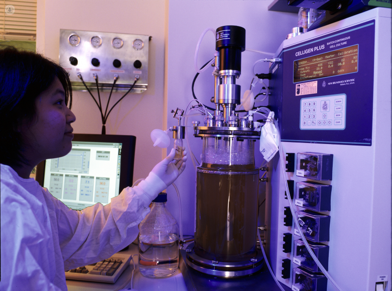
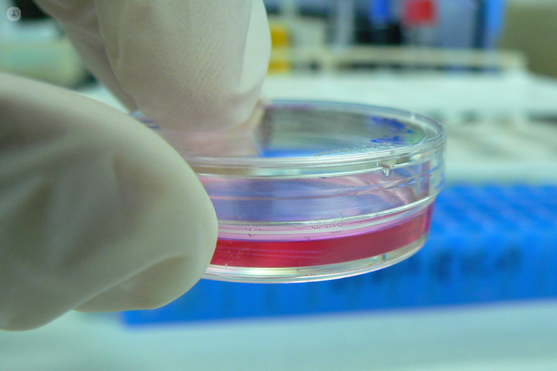
The best-known study is one that determines the foetal karyotype (an organised profile of a person’s chromosomes). A cell culture is carried out (the cells are grown in an artificial environment) for between 14 and 21 days, the chromosomes are observed and their number is studied to check if they present any anomaly. This is the time required to fully visualise all chromosomes.
A quicker study, although more limited, may be done if clinical or personal circumstances require. There are two techniques: FISH (Fluorescence in situ hybridisation) or QF-PCR (Quantitative Fluorescence-Polymerase Chain Reaction).
These techniques only study the numbers of certain chromosomes present in the foetal cells in the amniotic fluid. It does not study the whole chromosome; only the count. For example, we can check how many copies of chromosome 21 a foetal cell has: most people have two, but if there are three, this is what causes Down’s syndrome.
The advantage of this technique is that the result is known very quickly (24-48 hours), since it does not require the culture of the cells, but the disadvantage is that it does not detect all chromosomal abnormalities (it only detects the number of the studied chromosomes), so it is also necessary to perform the culture that takes 14-21 days, to obtain the complete foetal karyotype. In many clinics, both techniques are performed with the same sample.
As well as studying the chromosomes, the presence or production of certain substances by the foetus, or infections can be detected in the amniotic fluid.
Can a baby be born with genetic diseases if the karyotype is normal?
If the karyotype taken during amniocentesis is normal, it does not mean that the foetus definitely won’t have a genetic disease. Some genetic conditions are caused by specific genes, and there are thousands of genes on each chromosome, making it difficult to identify some of them from samples.
There are currently techniques (e.g. array CGH – comparative genomic hybridisation) that are able to determine in the amniotic fluid the genetic abnormalities that cause a group of serious conditions that would still present a normal karyotype (the anomaly would be in one of the thousands of genes of a chromosome). In addition, several hundred of them can be studied at the same time.
When is amniocentesis recommended?
In many medical centres, prenatal screening protocols have been put in place with the aim of reducing the number of amniocentesis studies so that amniocentesis would only be performed on those patients determined as high risk. This is because amniocentesis is invasive and has a small risk of miscarriage, so the procedure should only be carried out on patients with an increased risk of genetic and chromosomal conditions.
If you are considering this procedure, you should consult your doctor or a specialist.